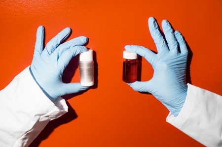
Detectarea cu ochi liber a vaporilor de solvenți(Naked-eye detection of solvent vapor)

a dezvoltat recent un senzor fluorescent eficient pentru vaporii de solvent. Senzorul lor se bazează pe o moleculă ramificată numită dendrimer. Dendrimerul este compus dintr-un nucleu de acceptare a electronilor atașat ramurilor donatoare de electroni. În anumite condiții, dendrimerul se auto-asamblează în fibre care formează un cadru cristalin care conține numeroase pori, ceea ce a ajutat la adsorbția moleculelor de solvent .demonstrează că senzația vizibilă de vapori de solvent poate fi realizată folosind dendrime cristaline cu structuri poroase. Astfel de dendrimeri arată promisiune pentru utilizarea în discriminarea gazelor și a materialelor organice, făcându-le atractive pentru aplicațiile de detectare a mediului.

Posted: Feb 28, 2018

Naked-eye detection of solvent vapor

(Nanowerk News) Sensors are important for detecting contaminants and monitoring environmental conditions. Fluorescent sensors can reveal the presence of a contaminant according to the change of the intensity and/or wavelength of light that they emit. In particular, fluorescent sensors that display changes in emission color are attractive because such changes may be readily observed with the naked eye, making them easy to use.

A research team led by the University of Tsukuba has recently developed an effective fluorescent sensor for solvent vapor. Their sensor is based on a branched molecule called a dendrimer. The dendrimer is composed of an electron-accepting core attached to electron-donating branches. Under certain conditions, the dendrimer self-assembled into fibers that formed a crystalline framework containing numerous pores, which aided adsorption of solvent molecules (Chemical Communications, „A fluorescent microporous crystalline dendrimer discriminates vapour molecules”).
Turn-On PL Sensor for Vapors
Turn-On PL Sensor for Vapors. (Image: University of Tsukuba)
“Slow diffusion of methanol or acetonitrile into chloroform solutions of the dendrimer yielded crystalline microfibers with high porosity,” states Sae Nakajima. “Under other conditions, we obtained amorphous microspheres of the dendrimer that lack pores.”
The research group investigated the crystal structure of the dendrimer by diffraction techniques, finding that the pore volume of the microfiber structure was over 70% of the total crystal volume. The high pore volume of the crystalline dendrimer microfibers greatly increased their gas adsorption capacity over that of the spheres formed under other conditions.
Films of the crystalline dendrimer displayed favorable emission behavior for use in solvent sensing. When the films were exposed to solvent vapor, their emission intensity increased considerably and their emission color quickly changed (within two seconds), reflecting the solvent polarity. This means that a single film could be used to detect numerous types of solvents.
For example, a film appeared blue in a fluorescence microscopy image before solvent exposure and became yellow after introduction of water vapor or green after methanol addition. The film returned to its original color when the solvent vapor was removed, allowing it to be reused.
“Dendrimers typically possess dense amorphous structures. The porous crystalline structure formed by our dendrimer under specific conditions increased its adsorption capacity and sensing ability, revealing the importance of crystalline structure in fluorescent sensing by dendrimers,” says Yohei Yamamoto.
This work demonstrates that the visible sensing of solvent vapor can be realized using crystalline dendrimers with porous structures. Such dendrimers show promise for use in discrimination of gases and organic materials, making them attractive for environmental sensing applications.
Source: University of Tsukuba

Pielea pieptului poate fi soluția pentru camuflarea materialului(Squid skin could be the solution to camouflage material)

Organele de cromatopor, care apar ca sute de pistrui multi-colorate pe suprafața corpului cefalopodului, contribuie la schimbări rapide ale culorii pielii.Cercetatorii au descoperit ca aceste granule au calitati optice remarcabile si le-au folosit pentru a face filme subtiri si fibre care ar putea fi incorporate in textile, afise flexibile si dispozitive viitoare de schimbare a culorii.construiasca aceste dispozitive care sa se potriveasca cu culoarea, schimbarea culorii si camuflajul la fel ca cefalopodii, dar multi dintre ei nu vin aproape de viteza sau gama dinamica de culoare pe care animalele le pot expune .

Posted: Feb 27, 2018

Squid skin could be the solution to camouflage material

(Nanowerk News) Cephalopods – which include octopuses, squid, and cuttlefish – are masters of disguise. They can camouflage to precisely match their surroundings in a matter of seconds, and no scientist has quite been able to replicate the spectacle. But new research by Leila Deravi, assistant professor of chemistry and chemical biology at Northeastern, brings us a step closer.

The chromatophore organs, which appear as hundreds of multi-colored freckles on the surface of a cephalopod’s body, contribute to fast changes in skin color. In a paper published in Advanced Optical Materials („Natural Light-Scattering Nanoparticles Enable Visible through Short-Wave Infrared Color Modulation”), Deravi’s group describes its work in isolating the pigment granules within these organs to better understand their role in color change.
The researchers discovered these granules have remarkable optical qualities and used them to make thin films and fibers that could be incorporated into textiles, flexible displays, and future color-changing devices. Deravi’s lab collaborated with the U.S. Army Natick Soldier Research, Development, and Engineering Center for the study.
Researcher Holding Spools of Thread Created From Squid Pigment
Northeastern researcher Leila Deravi displays spools of thread that were created using squid pigment. (Image: Adam Glanzman/Northeastern University)
Chromatophores come in shades of red, yellow, brown, and orange. They are similar to the freckles on human skin that appear over time. But in cephalopods, these freckles open and close within a fraction of a second to give rise to a continuously reconfiguring skin color. Underneath the chromatophores is a layer of iridophores that act as a mirror. Together, these organs reflect all colors of visible light.
By removing individual pigment particles from the squid, Deravi was able to explore the breadth of their capabilities as static materials. One particle is only 500 nanometers in size, which is 150 times smaller than the diameter of a human hair. Deravi’s team layered and reorganized the particles and found they could produce an expansive color pallet.
„We’re showing these pigments are a powerful tool that can produce ultra-thin films that are really rich in colors,” Deravi said.
Her team also discovered the pigments can scatter both visible and infrared light. This enhances brightness and light absorption and affects how a final color is perceived. And when Deravi engineered a system that included a mirror – mimicking the layout of organs that squids have naturally–she was able to further enhance the perceived color through scattering light through and off the granules. This process could potentially be replicated on functional materials like solar cells to increase the absorption of sunlight, Deravi said.
„From a scientific and technical engineering perspective, understanding how light scattering affects color is very important, and this is an exciting new development in the field of optics in biology,” said Richard Osgood, a collaborator from the U.S. Army Natick Soldier Research, Development, and Engineering Center. „This is an unusual harnessing of optics and physics knowledge in scattering to understand biological systems.”
The researchers made spools of fibers from the squids’ pigment particles and are now exploring uses for the material. The fibers are so visually interesting that it’s not difficult to imagine weaving them into fabric for clothing or other art forms. But perhaps the most exciting possible application is wearable, flexible screens and textiles that are capable of adaptive coloration. Osgood said the research could allow the Army to create new capabilities for soldiers.
„For more than a decade, scientists and engineers have been trying to replicate this process and build these devices that can color match, color change, and camouflage just like the cephalopods, but many of them come nowhere near the speed or dynamic range of color that the animals can display,” Deravi said. „Cephalopods have evolved to incorporate these specific pigment granules for a reason, and we’re starting to piece together what that reason is.”
Source: By Allie Nicodemo, Northeastern University

Emisiile optice ale sulfurii arsenice bidimensionale preparate în plasmă(Optical emission of two-dimensional arsenic sulfide prepared in plasma)

Datorită proprietăților sale, sulfura de arsenic, structurată în două straturi, structurată în două straturi, este un material promițător pentru dezvoltarea dispozitivelor fotosensibile 2D. Aceste proprietăți sunt, de asemenea, utile atunci când se creează tranzistoare cu efect de câmp, fotodetectoare cu sensibilitate ridicată și senzori de gaz.

Posted: Feb 27, 2018

Optical emission of two-dimensional arsenic sulfide prepared in plasma

(Nanowerk News) Since the discovery of graphene in 2004, there has been a rapidly growing interest among scientists in the study of 2D materials „beyond graphene”. In the family of chalcogenide materials, 2D-layered transition-metal dichalcogenides demonstrate excellent electronic and optical properties, outstanding mechanical flexibility, and exceptional catalytic performance. At the same time, chalcogenides like As2S3, As2Se3, etc., have never been considered as materials capable of forming structures of this type.

It could be due to the limitations of the methods used for preparing those materials. A group of researchers from the laboratory of functional nanomaterials at the Lobachevsky State University of Nizhny Novgorod, the Alekseev Nizhny Novgorod State Technical University and the AGH University of Science and Technology (Krakow, Poland) show the possibility of forming honeycomb structural fragments in plasma using the example of the well-known arsenic sulfide chalcogenide system (Spectrochimica Acta Part A: Molecular and Biomolecular Spectroscopy, „Infrared and Raman spectroscopy study of As-S chalcogenide films prepared by plasma-enhanced chemical vapor deposition”).
For the first time, optical emission of two-dimensional „beyond graphene” arsenic sulfide prepared in plasma has been demonstrated. A strong structural photoluminescence exited by continuous wave operation lasers with a laser excitation wavelength of 473 nm and 632.8 nm has been observed.
The influence of excitation parameters, chemical composition, structure, and annealing conditions on the intensity of photoluminescence of the chalcogenide materials has been established. Mass-spectrometry and Raman spectroscopy were coupled with quantum-chemical calculations to reveal the fragments which are the building blocks of the 2D As-S materials.
SEM Images of Arsenic Sulfide Materials
Figure 1. SEM images of arsenic sulfide materials prepared by plasma discharge at different conditions: pole-like (a) and layered (b) structures. (Image: Lobachevsky University) (click on image to enlarge)
A plausible mechanism of formation and modification of the arsenic sulfide luminescent structural units has been proposed. The properties of 2D pole-structured and layered arsenic sulfide are suggested to be the key to advancing to 2D photosensitive devices.
Scanning Electron Microscopy (SEM) images typical of the arsenic sulfide with a composition As40S60 are illustrated in Figure 1 (a) and (b). The striking difference in the surface morphology and structure is due to very different conditions of plasma deposition.
Both pictures (1a and 1b) illustrate arsenic sulfide structures consisting of (As2S2)n-units (see below) formed in plasma by spherical structural fragments with a diameter of about 100 nm. Figure 1a shows a pole-structured material and Figure 1b depicts a 2D layered structure, the theoretical possibility of existence of which has been described recently.
The researchers have reported quantum-chemical estimations of these structures together with experimental results on the unusually broad transparency window of these materials (0.43 – 20 µm) in comparison with those of As2S3 (0.6 – 11 µm) prepared by traditional thermal methods.
Photoluminescence of the plasma prepared arsenic sulfide was measured by excitation at 473 nm and 632.8 nm employing continuous wave operation lasers at room temperature (RT, Figure 2).
Photoluminescence and Mass Spectra of the Plasma Prepared Arsenic Sulfide
Figure 2. This is a photoluminescence and mass spectra of the plasma prepared arsenic sulfide. (Image: Lobachevsky University) (click on image to enlarge)
The arsenic sulfide materials prepared in plasma were analyzed using the mass-spectrometry method to reveal and to gain insight into the main structural fragments affecting the luminescence intensity.
The mass-spectroscopy data presented clarify the main structural fragments affecting the PL intensity of the plasma prepared arsenic sulfide. According to the data obtained and taking into account the point of view formulated previously we may assume that the main reason for appearance and enhancing of the luminescence in arsenic sulfide materials prepared in plasma is the (As2S2)n cyclic structure unit playing the role of a „disk-like polarizability tensor”.
Due to its properties, two-dimensional pole-structured and layered „beyond graphene” arsenic sulfide is a promising material for developing 2D photosensitive devices. These properties are also useful when creating field-effect transistors, highly sensitive photodetectors and gas sensors.
Source: Lobachevsky University

Wilele lui E. Coyote inspiră dezvoltarea unor noi proprietăți ale siliciului(Wile E. Coyote’s fall inspires development of new properties of silicon)

Rezultatele arată că siliciul poate avea un nou contract de închiriere a vieții, oferind noi modalități de a controla informațiile cu lumină, mai degrabă decât cu curent electric, ceea ce înseamnă computere mult mai rapide și comunicații cu o lățime de bandă mai mare

Posted: Feb 27, 2018

Wile E. Coyote’s fall inspires development of new properties of silicon

(Nanowerk News) An international team of scientists, led by the University

of Surrey, has discovered a new type of silicon that could be used to control light beams in a new kind of photonic chip – a chipset where information is carried by light beams rather than electrical currents.

The essence of the technology – where an object takes a moment to respond to the energy placed upon it – is a staple of cartoons such as Roadrunner, where characters run off cliffs and spend a moment in mid-air before falling.

Scientists hope that their discovery, detailed in a study published by Nature Photonics („Giant multiphoton absorption for THz resonances in silicon hydrogenic donors”), will lead to the development of more exciting technologies such as signal modulators for terahertz (THz) beams – which is part of the electromagnetic spectrum between visible/infrared light and radio/microwaves.

Silicon is widely used to send microwave signals for mobile communications, but it is very poor at sending visible light signals. The team discovered that the standard impurities that are sprinkled into ordinary computer chips to make transistors can control the flow of THz photons far more efficiently than almost anything else. This has the double benefit of potentially allowing a new method of chip-to-chip communication with silicon, currently only possible with much more expensive materials, but also pushing mobile communications to much higher frequency and allowing the transmission of more data.

The signal modulation effect works by using two or more photons, each of which could individually go straight through the silicon unhindered, and only when they arrive together they get absorbed. The first photon acts like a switch – its presence or absence determines what will happen to the others. The catch is that the second photon has to be almost simultaneous with the first, meaning that the intensity of the beams must be really high. The researchers tried using THz photons instead of the infrared photons used in all previous attempts, and found that they could get switching with thousands of times lower intensity than ever before.

Professor Ben Murdin from the University of Surrey, said: “It’s just like when Wile E. Coyote is chasing the Roadrunner and goes off the edge of a cliff – there’s always a moment before physics wakes up and realises he has too much potential energy and he falls. During this ‘coyote time’ (as gamers call it) sometimes something else can take effect like a rocket or a stone or a jump. That’s exactly how Heisenberg’s Uncertainty Principle works here – there’s a little bit of ‘coyote time’ after the first photon hits in which the molecule doesn’t know what energy it’s supposed to have, but the more energy it tries to ignore the less the coyote time available.

“We found that with terahertz light silicon’s coyote time is much, much longer, meaning this kind of photon switch is far more efficient than anything else we know of. The results show that silicon may have a completely new lease of life, providing new ways to control information with light rather than electrical current, meaning far faster computers and higher bandwidth communications.”

Source: University of Surrey

Proprietățile optice ale materialelor 2D ar putea avansa tehnologia infraroșie(Optical properties of 2D materials could advance infrared technology)

au studiat caracteristicile optice ale unui tip special de material fabricat dintr-un singur strat de atomi de fosfor in folosul detectiei si interactiunii cu lumina infrarosie, invizibil nu numai pentru ochiul uman, ci si pentru multi alte materiale propuse pentru utilizare în sistemele optoelectronice.Studiul a avansat, de asemenea, înțelegerea naturii plasmonice a acestui material atunci când este modelat în panglici nanometrice cu diferite lățimi. Combinând parametrii de reglare a lățimii nanoribbonului și a mediului înconjurător, fosforul negru poate deveni un material foarte util pentru aplicațiile care implică lumină infraroșie.
Un avantaj suplimentar al fosforului negru este faptul că cele două direcții cristaline diferite permit ca lumina să interacționeze diferit cu electronii de pe suprafață, în funcție de orientarea undelor luminoase utilizate în aplicație.

Posted: Feb 27, 2018

Optical properties of 2D materials could advance infrared technology

(Nanowerk News) University of Arkansas researchers have studied the optical characteristics of a special type of material made of a single layer of phosphorus atoms for the benefit of detecting and interacting with infrared light, which is invisible not only to the human eye but to many other materials proposed for use in optoelectronic systems.

Such systems strive to utilize light alongside electrons to speed up processing and to reduce heating and other energy-wasters in our ever-growing plethora of computational devices.
The group of researchers in the Department of Physics published their findings in a recent issue of Scientific Reports („Tuning Infrared Plasmon Resonance of Black Phosphorene Nanoribbon with a Dielectric Interface”), helping to advance the understanding of black phosphorus as an optically useful material. Physics doctoral student Desalegn Debu was the first author of this theoretical and computational work. Other authors include Stephen Bauman and David French, University of Arkansas graduate students; and Hugh Churchill and Joseph Herzog, assistant professors in the Department of Physics.
Similar to a pebble splash causing waves to move across the surface of a pond, light shining upon a plasmonic material causes electrons to move back and forth on the surface. This wave of electrons, known as a plasmon, can be tuned like a musical instrument to oscillate most strongly for certain frequencies (colors) of light, making them useful for applications ranging across much of the spectrum of visible and invisible signals. Tuning is key to the use of plasmonic materials for specific applications, just as tuning a musical instrument is critical to creating the desired note.
Two-dimensional materials such as graphene, a sheet of carbon with single-atom thickness, have experienced a lot of hype in the scientific world within the past decade. They offer the potential of reducing the size of various optoelectronic components down to single atoms thick as well as taking advantage of unique physical properties.
While graphene has been the ‘poster child’ of 2D materials and it may yet hold promise for improved electronics, material strength, or light-based technology, it is not perfect. Graphene lacks what is called a bandgap, a defining property of semiconductors. This limitation of graphene is being circumvented by looking into other materials that offer superior structural, electrical, thermal, or optical properties.
Black phosphorus, which has a corrugated structure of phosphorus atoms with two different crystalline directions, offers unique benefits over other previously studied options.
The study performed by Debu and his colleagues investigates the theoretical effects of changing material properties in the area surrounding a black phosphorus sheet. The results demonstrate that the wavelength of light absorbed by the material can be tuned by changing the surrounding material.
The study also advanced the understanding of the plasmonic nature of this material when it is patterned into nanoscale ribbons with different widths. Combining the tuning parameters of nanoribbon width and surrounding media, black phosphorus can become a very useful material for applications involving infrared light.
An additional benefit of black phosphorus is that its two different crystalline directions allow for light to interact differently with the electrons on the surface depending on the orientation of the light waves used in the application.
Source: University of Arkansas

Nanocoarea inteligentă deschide ușa spre ferestrele inteligente(Clever nanocoating opens door to smart windows)

au dezvoltat un nou strat ultra-subțire care răspunde la căldură și frig, deschizând ușa spre „ferestrele inteligente”.

Vopseaua de auto-modificare, care este de o mie de ori mai subțire decât un fir de păr uman, funcționează automat, permițându-i să se încălzească automat când este rece și blocând razele soarelui atunci când este fierbinte.

Ferestrele inteligente au capacitatea de a regla în mod natural temperaturile din interiorul unei clădiri, ceea ce duce la beneficii majore pentru mediu și la economii semnificative.

Posted: Feb 26, 2018

Clever nanocoating opens door to smart windows

(Nanowerk News) Researchers from RMIT University in Melbourne Australia have developed a new ultra-thin coating that responds to heat and cold, opening the door to „smart windows”.

The self-modifying coating, which is a thousand times thinner than a human hair, works by automatically letting in more heat when it’s cold and blocking the sun’s rays when it’s hot.

Smart windows have the ability to naturally regulate temperatures inside a building, leading to major environmental benefits and significant financial savings.

Lead investigator Associate Professor Madhu Bhaskaran said the breakthrough will help meet future energy needs and create temperature-responsive buildings.

Mohammad Taha shows off an ultra-thin coating

Mohammad Taha shows off the ultra-thin coating developed at RMIT University. (Image: RMIT University)

„We are making it possible to manufacture smart windows that block heat during summer and retain heat inside when the weather cools,” Bhaskaran said.

„We lose most of our energy in buildings through windows. This makes maintaining buildings at a certain temperature a very wasteful and unavoidable process.

„Our technology will potentially cut the rising costs of air-conditioning and heating, as well as dramatically reduce the carbon footprint of buildings of all sizes.

„Solutions to our energy crisis do not come only from using renewables; smarter technology that eliminates energy waste is absolutely vital.”

Smart glass windows are about 70 per cent more energy efficient during summer and 45 per cent more efficient in the winter compared to standard dual-pane glass.

New York’s Empire State Building reported energy savings of US$2.4 million and cut carbon emissions by 4,000 metric tonnes after installing smart glass windows. This was using a less effective form of technology.

„The Empire State Building used glass that still required some energy to operate,” Bhaskaran said. „Our coating doesn’t require energy and responds directly to changes in temperature.”

Co-researcher and PhD student Mohammad Taha said that while the coating reacts to temperature it can also be overridden with a simple switch.

„This switch is similar to a dimmer and can be used to control the level of transparency on the window and therefore the intensity of lighting in a room,” Taha said. „This means users have total freedom to operate the smart windows on-demand.”

Windows aren’t the only clear winners when it comes to the new coating. The technology can also be used to control non-harmful radiation that can penetrate plastics and fabrics. This could be applied to medical imaging and security scans.

Bhaskaran said that the team was looking to roll the technology out as soon as possible.

„The materials and technology are readily scalable to large area surfaces, with the underlying technology filed as a patent in Australia and the US,” she said.

The research has been carried out at RMIT University’s state-of-the-art Micro Nano Research Facility with colleagues at the University of Adelaide and supported by the Australian Research Council.

Their findings have been published in Scientific Reports („Insulator–metal transition in substrate-independent VO2 thin film for phase-change devices”).

How the coating works

The self-regulating coating is created using a material called vanadium dioxide. The coating is 50-150 nanometres in thickness.

At 67 degrees Celsius, vanadium dioxide transforms from being an insulator into a metal, allowing the coating to turn into a versatile optoelectronic material controlled by and sensitive to light.

The coating stays transparent and clear to the human eye but goes opaque to infra-red solar radiation, which humans cannot see and is what causes sun-induced heating.

Until now, it has been impossible to use vanadium dioxide on surfaces of various sizes because the placement of the coating requires the creation of specialised layers, or platforms.

The RMIT researchers have developed a way to create and deposit the ultra-thin coating without the need for these special platforms – meaning it can be directly applied to surfaces like glass windows.

Source: RMIT University

Cercetătorii găsesc metale în catalizatori grafeni „fără metale”(Researchers find metal in ‘metal-free’ graphene catalysts)

Grafenul a fost testat pe scară largă ca înlocuitor pentru platina scumpe în aplicații precum celulele de combustie, unde materialul catalizează reacția de reducere a oxigenului (ORR) esențială pentru transformarea energiei chimice în energie electrică. Trei imagini microscopice cu transmisie electronică cu grafeni dopat cu azot arată prezența relativă a atomilor de mangan.Catalizatorii cu un atom atomic se pot ascunde printre grafen și activitatea lor este profundă”, a spus el. „Ceea ce a fost uneori atribuit grafenului a fost într-adevăr singurul metal îngropat în suprafața grafenului. Graphenul este bun în sine, dar în aceste cazuri, a fost făcut să arătă chiar mai bine de acești atentatori cu un singur atom de metal.Catalizatorii cu un atom atomic se pot ascunde printre grafen și activitatea lor este profundă”, a spus el. „Ceea ce a fost uneori atribuit grafenului a fost într-adevăr singurul metal îngropat în suprafața grafenului. Graphenul este bun în sine, dar în aceste cazuri, a fost făcut să arătă chiar mai bine de acești atentatori cu un singur atom de metal.

Posted: Feb 26, 2018

Researchers find metal in ‘metal-free’ graphene catalysts

(Nanowerk News) Detective work by Rice University chemists has defined a deception in graphene catalysts that, until now, has defied description.

Graphene has been widely tested as a replacement for expensive platinum in applications like fuel cells, where the material catalyzes the oxygen reduction reaction (ORR) essential to turn chemical energy into electrical energy. Three transmission electron microscope images of nitrogen-doped graphene show the relative presence of manganese atoms

Three transmission electron microscope images of nitrogen-doped graphene show the relative presence of manganese atoms, contaminants from graphite precursors or reactants believed responsible for the material’s ability to catalyze oxygen-reduction reactions, according to Rice University scientists. The top image shows many manganese atoms (white) remain on graphene that has been washed once; few on twice-washed graphene in the center image; and none on graphene washed six times at bottom. Twice-washed graphene with a scattering of manganese atoms proved best for catalysis. (Image: Tour Group/Rice University)Because graphene, the atom-thick form of carbon, isn’t naturally metallic, researchers have been baffled by its catalytic activity when used as a cathode.


Wonder no more, said Rice chemist James Tour and his crew, who have discovered that trace quantities of manganese contamination from graphite precursors or reactants hide in the graphene lattice. Under the right conditions, those metal bits activate the ORR. Tour said they also provide insight into how ultrathin catalysts like graphene can be improved.


The research appears in the journal Carbon („Manganese deception on graphene and implications in catalysis”).


Because the contrast between carbon and manganese atoms is so slight, trace atoms of the contaminants can’t be seen with traditional characterization techniques like X-ray diffraction and X-ray photoelectron spectroscopy (XPS).


“Labs have reported ‘metal-free’ graphene catalysts, and the evidence they’ve gathered could easily be interpreted to show that,” Tour said. “In fact, the tools they were using simply weren’t sensitive enough to show the manganese atoms.”


A more sensitive tool, inductively coupled plasma mass spectrometry (ICP-MS), clearly saw the interlopers among samples made by the Rice lab.


Nitrogen-doped graphene test samples were reduced from graphene oxide and then acid-washed between one and six times. With each wash, the ICP-MS scan showed fewer manganese atoms and detected none in graphene samples washed six times. By the fifth wash, the catalytic activity totally changed and showed the former activity had been due to those residual metal atoms.


The lab reported no manganese atoms were observed in any of the same samples using conventional analytical tools, including XPS or transmission electron microscopy.


The researchers characterized the samples’ ORR activity and found twice-washed nitrogen-graphene was most effective. These samples tended to incorporate single atoms of manganese into the graphene structure, which facilitated full reduction of oxygen through a four-electron process in which four electrons are transferred to oxygen atoms, usually from hydrogen.


“In a four-electron process, oxygen is reduced to water or hydroxide,” said Rice graduate student Ruquan Ye, the paper’s lead author. “However, peroxide is formed in a two-electron process, which results in a lower diffusion-limited current density and generates hazardous reactive oxygen species.” Ye said that without metal, the ORR in graphene is far less efficient.


Tour said the results should lead to investigation of the role of trace metals in other materials thought to be metal-free.

“Single-atom catalysts can hide among graphene, and their activity is profound,” he said. “So what has sometimes been attributed to the graphene was really the single metal buried into the graphene surface. Graphene is good in its own right, but in these cases, it was being made to look even better by these single metal-atom stowaways.”

Source: Rice University

Firele din nanotuburi de carbon intaresc nervii pentru tratamentele electrochirurgie si diagnosticare(Carbon nanotube yarn taps nerves for electroceutical treatments and diagnostics)

Acum, cercetările finanțate de NIBIB au transformat nanotuburile de carbon în fire sau fire flexibile, de natură nervoasă, capabile de legături de lungă durată de înaltă fidelitate la animalele vii. Dezvoltarea acestor fire biocompatibile deschide posibilitatea unor noi diagnostice și terapii bioelectrice prin reglarea funcției interne a organelor la nivelul nervului unic.
Nanotuburile individuale de carbon sunt trase de pe un substrat și se rotesc în firele flexibile de nanotuburi de carbon

Posted: Feb 26, 2018

Carbon nanotube yarn taps nerves for electroceutical treatments and diagnostics

(Nanowerk News) Ingested or injected pharmaceuticals can target specific molecules involved in disease processes, but get distributed throughout the body where they can cause unwanted side effects. An approach known as electroceuticals aims to avoid systemic exposure by using small wires to electrically monitor and manipulate individual nerves that control organ function and carry information about disease. Despite the promise of electroceuticals, it has been challenging to develop long-term therapies due to the lack of biocompatible wires.

Now, NIBIB-funded researches have spun carbon nanotubes into flexible, nerve-sized wires or yarns capable of high-fidelity long-term connections in live animals. The development of these biocompatible yarns opens the possibility of new bioelectric diagnostics and therapies through regulation of internal organ function at the single nerve level.

Individual carbon nanotubes are pulled from a substrate and spun into the flexible carbon nanotube yarn

Individual carbon nanotubes are pulled from a substrate and spun into the flexible carbon nanotube yarn. (© NPG)

All the organs of the body such as the heart, lungs, liver, and kidneys are automatically controlled by nerves that stretch from the brainstem to each organ. These nerves control organ functions such as heartbeat, breathing rates, and blood pressure, making constant adjustments in response to environmental and physiological changes. Variations in the electrical activity in this area of the brain, known as the autonomic nervous system, can also be predictors, indicators or causes of disease development.

“Monitoring and manipulating the autonomic nervous system to both understand and potentially treat disease has been an intriguing yet understudied approach to medicine,” explains Michael Wolfson, Ph.D., director of the NIBIB program in Rehabilitation Engineering and Implantable Medical Devices. “This is largely due to technical limitations in being able to insert wires into nerves that can reliably and safely record electrical activity over long periods of time and under different physiological conditions.”

In a study reported in the journal Scientific Reports („Chronic interfacing with the autonomic nervous system using carbon nanotube (CNT) yarn electrodes”), bioengineers at Case Western Reserve University in Cleveland, Ohio describe the development of highly flexible carbon nanotube (CNT) yarn electrodes that were capable of months-long electrical recording in major nerves of the autonomic nervous system in rats.

The CNT yarns are essentially, just that, yarns made from a “forest” of hundreds of carbon nanotubes that are pulled from the metal surface they are grown on and spun into a highly flexible, highly conductive wire 1/100th the size of a human hair.

carbon nanotube yarn is wound around a tungsten needle for insertion into a nerve

The carbon nanotube yarn is wound around a tungsten needle for insertion into a nerve. The coiling of the yarn causes it to remain solidly embedded in the nerve after the needle is withdrawn. Actual needle and yarn (top). Diagram showing detail of how yarn is wrapped around the needle for insertion (bottom). (© NPG)

Current technologies for recording electrical signals from nerves include relatively large stiff tungsten needles used by neurologists to obtain readings from single nerves of patients for several hours, but must be removed before causing lasting nerve damage. Other wire electrode technologies can record nerve signals for short periods but because of their dimensions and stiff mechanical properties they are not suitable for long-term recording in small nerves.

“The currently available electrode technologies simply do not match the mechanical properties of nerves,” explains Dominique Durand, Ph.D., Professor of Biomedical Engineering at Case Western and senior researcher on the CNT yarn work. “With those types of electrodes, it’s often like sticking glass into spaghetti. Our CNT yarns are similar in size and flexibility to actual nerves. These properties allow them to be stealthily inserted into specific nerves and remain there for months without destroying the tissue or inducing an attack by the immune system.”

The combination of the biocompatibility of the CNT yarns and their outstanding ability to carry an electrical signal that is 10 times stronger than current technologies makes them ideal for long term recording of specific nerve signals. Finally, the lack of nerve damage keeps the surrounding axons intact, which helps to eliminate background noise. Thus, the CNT yarns have an excellent signal to noise ratio (SNR), which is critical for this type of research.

The group tested the CNT yarns in two major nerves in the autonomic system in rats. One study involved the vagus nerve, which stretches throughout the body to connect to numerous organs. The nerve is known to control and monitor a range of functions including heart rate, digestive tract movement, sweating, and immune response.

CNT yarn electrodes were also inserted into the glossopharyngeal nerve. The nerve is connected to a number of organs including the carotid artery, and parts of the ear, tongue and salivary glands where it is known to be involved in swallowing.

Carbon nanotube yarn embedded into the vagus nerve of a rat

Carbon nanotube yarn embedded into the vagus nerve of a rat. (© NPG)

In both nerves, recordings of stable electrical activity were maintained over a 10-week period. Pulses of nerve activity were also monitored while the animal responded to physiological challenges. The challenges included distension of the rat’s stomach with saline solution, and short durations where the rats were in low oxygen environments. In each case, the physiological changes induced by the challenge resulted in easily detectable changes in electrical activity that were recorded using the CNT yarn implants over the entire 10-week period of the experiments.

“Although this is early research, we believe the results demonstrate that this technology can be used for reliable long term electrical monitoring of physiological functions,” says Durand. “This is an important step in pursuing monitoring of disease progression through the nerves that control the function of the diseased organ.”

Durand explains that the goal is to learn what electrical signatures or profiles are indicative of disease development and to use that for early intervention, potentially by electrical stimulation or even blocking of the nerve. For example, one therapy for severe hypertension is the cutting of the renal nerve. This is obviously irreversible. Durand, explained that CNT yarn electrodes could be used to block the nerve impulse without cutting the nerve, making the treatment reversible.

The group is also excited about the potential of this recording method to improve prosthetics. “When an arm has been amputated there are tens of thousands of neurons remaining,” said Durand. “The approach would be to insert the CNT yarns into individual nerves and record the electrical signals that are created as the individual thinks about moving the missing arm—essentially learning the electrical signals that are formed by the intention to move the arm.” The ultimate goal is to develop neural interfaces capable of translating those electrical signals into better control of the prosthetic by the user.

Source: National Institute of Biomedical Imaging and Bioengineering

Primul nanometru arată cum ionii de litiu navighează într-un labirint molecular pentru a ajunge la electrodul bateriei(First nanoscale look at how lithium ions navigate a molecular maze to reach battery electrode)

cercetătorii au reușit să determine structurile și pozițiile individuale molecule de solvenți și ioni de litiu care se aflau la câteva miliarde de metru din suprafața electrodului, a declarat Hans-Georg Steinrück, cercetător postdoctoral în grupul lui Toney și co-conducător al experimentelor. Simularea dinamicii moleculare a fost completă și convenită cu rezultatele experimentale.

„Putem vedea pozițiile de ioni și molecule de solvenți în apropierea electrodului cu rezoluție angstrom și, de asemenea, să vedem cum sunt orientate la suprafața electrodului”, a spus Steinrück. „Sunt aranjate în straturi bine definite la limită, iar primul strat este plat, paralel cu suprafața electrodului; atunci ele devin mai dezordonate, mai tipice pentru un lichid, pe măsură ce vă deplasați de pe suprafață. „Aceste straturi comandate îngreunează ionii de litiu să se miște rapid prin straturi și în electrod.

Posted: Feb 26, 2018

First nanoscale look at how lithium ions navigate a molecular maze to reach battery electrode

(Nanowerk News) The lithium-ion batteries that power laptops, electric cars and so many other modern gadgets operate on a simple plan: Lithium ions shuttle back and forth between two electrodes, inserting themselves into one of the electrodes as the battery charges and moving across to the other as the battery drains. The speed and ease of their travel through the battery’s liquid electrolyte help determine how fast the battery can charge.

Now scientists have taken the first close look at what happens within a few nanometers of the electrode, where the normally freely moving electrolyte molecules organize themselves into layers that stand directly in the lithium ions’ paths.

They directly observed this layering for the first time in X-ray experiments at the Department of Energy’s SLAC National Accelerator Laboratory. The results suggest that changing the concentration of lithium ions in the electrolyte might change the arrangement of the molecular layers and make it easier for the ions to get in and out of the electrode.

An illustration of electrolyte molecules arranging themselves into layers within a few nanometers of a battery electrode

An illustration of electrolyte molecules arranging themselves into layers within a few nanometers of a battery electrode. Lithium ions (purple balls) must navigate through these layers on their way in and out of an electrode during battery charging and discharging. (Image: Greg Stewart/SLAC National Accelerator Laboratory)

“That process of the ions finding their way into the electrode is very important in terms of how fast you can charge the battery and how long the battery lasts,” said Michael Toney, a distinguished staff scientist at SLAC’s Stanford Synchrotron Radiation Lightsource (SSRL) and co-leader of the study. “Understanding the nanoscale details of how this works could suggest ways to increase charging speed and efficiency.”

The report has been accepted for publication in Energy & Environmental Science („The nanoscale structure of the electrolyte–metal oxide interface”), and an advance copy is postedon the journal’s website.

Probing a Commercial Electrolyte

In lithium-ion batteries, the electrolyte consists of lithium and other ions in a solvent, with the solvent molecules moving around as they would in any other liquid. But based on theory and previous computer simulations, scientists had a strong suspicion that something different happened in the tiny volume of the electrolyte that’s right up next to the electrode. Here, they thought, the presence of the electrode’s hard surface would induce the solvent molecules to line up and form orderly layers. However, confirming this through experiments proved difficult.

For these latest experiments, Toney’s team used a metal oxide material to represent the electrode, bathed in an electrolyte typically found in commercial lithium-ion batteries.

By focusing a high-brilliance X-ray beam from SSRL on the surface of the electrode and analyzing the X-rays that bounced back through the electrolyte, like light reflecting from a mirror, the researchers were able to determine the structures and positions of individual solvent molecules and lithium ions that were within a few billionths of a meter of the electrode surface, said Hans-Georg Steinrück, a postdoctoral researcher in Toney’s group and co-leader of the experiments. Molecular dynamics simulations complemented and agreed with the experimental results.

“We can see the positions of ions and solvent molecules near the electrode with angstrom resolution, and also see how they are oriented at the surface of the electrode,” Steinrück said. “They’re arranged in well-defined layers at the boundary, and the first layer lies flat, parallel to the surface of the electrode; then they become more disordered, more typical of a liquid, as you move out from the surface.” These ordered layers make it more difficult for the lithium ions to move quickly through the layers and into the electrode.

Shifting Ranks of Molecules

However, as the concentration of lithium ions in the electrolyte increased, the arrangement of the layers changed; it became a bit more orderly, and the layers were farther apart, Steinrück said. This led the researchers to a conclusion that seems almost the opposite of what you’d expect.

“Our hypothesis is that if you want to improve lithium ion transport, you want to decrease the amount of order in the layers, and that means decreasing the lithium ion concentration rather than increasing it,” he said.

Steinrück said the team will be exploring this avenue of research further, adding that the foundational knowledge obtained with this technique can also be applied to studies of other types of next-generation batteries and energy storage systems.

Source: By Glennda Chui, SLAC National Accelerator Laboratory


Uniforme inteligente de luptă pentru unități speciale IDF(Smart Combat Uniform to IDF Special Units)

Mutarea inovării a fost recent raportată în domeniul uniformei inteligenței.Noua uniformă de luptă din materiale avansate, care va fi livrată în curând către unitățile speciale IDF, va îmbunătăți eficiența operațională și performanța de luptă pe termen lung și va spori supraviețuirea forțelor.  Uniformele vor fi montate pe echipamentul de luptă și vor fi confecționate dintr-un material rezistent la foc, rezistent la foc și sudoare

Smart