
Proiectarea implanturilor medicale disolubile(Designing Dissolvable Medical Implants)
Aceste implanturi sunt foarte personalizabile, astfel încât acestea pot fi modificate pentru a se potrivi nevoilor specifice ale fiecărui pacient
Aceste implanturi sunt foarte personalizabile, astfel încât acestea pot fi modificate pentru a se potrivi nevoilor specifice ale fiecărui pacient
Cu toate acestea, datorită răspândirii în creștere a bacteriilor cu rezistență multiplă, rezervorul antibiotic actual pare să fie insuficient, ceea ce necesită explorarea unor noi agenți antibacterieni
Departamentul de Apărare al SUA are un interes special în depășirea bolilor infecțioase .Membrii serviciului militar sunt chemați să opereze practic oriunde în lume, de multe ori pe termen scurt, iar locațiile pe care le desfășoară în mod frecvent nu dispun de publicul robust
Biologii din campusul din Florida al Institutului Scripps Reseaech au descoperit un nou mecanism care probabil stă la baza felului în care simțim forța sau atingerea
Progrese semnificative in tratamentele medicale de rani severe de arsuri, infectii si septicemii ulterioare persistente , cauze frecvente de morbiditate si mortalitate pentru victimele arsurilor.
Cercetatorii de la Universitatea Queen Mary din Londra au dezvoltat un nou mod de a dezvolta materiale mineralizate care ar putea regenera tesuturile tari, cum ar fi smaltul si osul dentar.Materialele ar putea fi utilizate pentru o mare varietate de complicații dentare, cum ar fi prevenirea și tratamentul cariilor dentare sau sensibilității la dinți – cunoscută și sub numele de hipersensibilitate la dentină.
| Posted: Jun 01, 2018 | |
Scientists develop material that could regenerate dental enamel(Nanowerk News) Researchers at Queen Mary University of London have developed a new way to grow mineralised materials which could regenerate hard tissues such as dental enamel and bone. |
|
| Enamel, located on the outer part of our teeth, is the hardest tissue in the body and enables our teeth to function for a large part of our lifetime despite biting forces, exposure to acidic foods and drinks and extreme temperatures. This remarkable performance results from its highly organised structure. | |
| However, unlike other tissues of the body, enamel cannot regenerate once it is lost, which can lead to pain and tooth loss. These problems affect more than 50 per cent of the world’s population and so finding ways to recreate enamel has long been a major need in dentistry. | |
| The study, published in Nature Communications („Protein disorder–order interplay to guide the growth of hierarchical mineralized structures”), shows that this new approach can create materials with remarkable precision and order that look and behave like dental enamel. | |
 |
|
| Close-up of the enamel-like material. (Image: Alvaro Mata) | |
| The materials could be used for a wide variety of dental complications such as the prevention and treatment of tooth decay or tooth sensitivity – also known as dentin hypersensitivity. | |
| Dr Sherif Elsharkawy, a dentist and first author of the study from Queen Mary’s School of Engineering and Materials Science, said: „This is exciting because the simplicity and versatility of the mineralisation platform opens up opportunities to treat and regenerate dental tissues. For example, we could develop acid resistant bandages that can infiltrate, mineralise, and shield exposed dentinal tubules of human teeth for the treatment of dentin hypersensitivity.” | |
| The mechanism that has been developed is based on a specific protein material that is able to trigger and guide the growth of apatite nanocrystals at multiple scales – similarly to how these crystals grow when dental enamel develops in our body. This structural organisation is critical for the outstanding physical properties exhibited by natural dental enamel. | |
| Lead author Professor Alvaro Mata, from Queen Mary’s School of Engineering and Materials Science, said: „A major goal in materials science is to learn from nature to develop useful materials based on the precise control of molecular building-blocks. The key discovery has been the possibility to exploit disordered proteins to control and guide the process of mineralisation at multiple scales. Through this, we have developed a technique to easily grow synthetic materials that emulate such hierarchically organised architecture over large areas and with the capacity to tune their properties.” | |
| Enabling control of the mineralisation process opens the possibility to create materials with properties that mimic different hard tissues beyond enamel such as bone and dentin. As such, the work has the potential to be used in a variety of applications in regenerative medicine. In addition, the study also provides insights into the role of protein disorder in human physiology and pathology. |
| Source: Queen Mary University of London |
Biosenzorii bazați pe puncte cuantice de siliciu pot fi utilizați ca un instrument de diagnosticare precoce pentru diferite afecțiuni, în timp ce nanocristalele de siliciu prezintă un mare potențial pentru dezvoltarea dispozitivelor de emisie de lumină bazate pe siliciu și producerea celulelor solare foarte eficiente.
| Posted: Apr 09, 2018 | |
Gold changes the photoluminescence of silicon quantum dots(Nanowerk News) A group of scientists from Russia and Sweden have demonstrated that applying gold stripes to a sample with silicon quantum dots modifies the dots’ properties. Their results of the study were published in Scientific Reports („Plasmon induced modification of silicon nanocrystals photoluminescence in presence of gold nanostripes”). |
|
 |
|
| (Image: Sergey Dyakov, Nikolay Gippius) (click on image to enlarge) | |
| Silicon quantum dots have a wealth of potential applications in cell biology and medicine thanks to their selective cell penetration capability and luminescence. Biosensors based on silicon quantum dots can be used as an early diagnosis tool for various diseases, while silicon nanocrystals hold great potential for developing silicon-based light-emitting devices and producing highly efficient solar cells. | |
| Scientists from the theoretical nanophotonics team led by Skoltech Professor Nikolay Gippius jointly with researchers from Moscow State University and the Royal Institute of Technology (KTH) in Stockholm demonstrated, first theoretically and then experimentally, that gold nanostripes are capable of changing the extinction and photoluminescence spectra of silicon nanocrystals. They established that the nanostripe width also affects the optical properties of the quantum dots. | |
| The researchers’ findings are of particular value for practical applications of silicon nanocrystals, as they provide scientists with a novel tool for controlling the quantum dots’ properties. | |
| Interestingly, the use of silicon quantum dots in photocells would significantly reduce technical losses in the solar power industry and help to avoid using arsenic, lead and other toxicants in solar cells production. | |
| “Derived from the interaction between a sample of silicon quantum dots and gold nanostripes is a quasiparticle called the ‘waveguide plasmon polariton.’ This leads to a change in the optical properties of silicon nanocrystals. If we wish to use them in solar batteries or light emitters, we should have a metal electrode and know how to control the extinction and emission spectra. | |
| Gold nanostripes are a solution to both issues,” said Sergey Dyakov, the main author of the study and a Skoltech researcher. |
| Source: Skolkovo Institute of Science and Technology |
Decuparea fantelor în stil kirigami în filme elastice ar putea face pentru bandaje, plăcuțe de căldură și electronice care pot fi purtate pe suprafețe flexibile
Prezentăm un citometru de impedanță microfluidică care poate fi purtat pe bandă flexibilă cu cititor de smartphone on-line pentru numărarea și analiza portabilă a biomarkerilor
Noile nanoparticule dezvoltate de Zibiao Li de la Institutul de Materiale de Cercetare și Inginerie A * STAR și colaboratorii săi, reprezintă un upgrade semnificativ față de generațiile anterioare de nanoparticule de eliberare a medicamentelor pe bază de polimeri. Exemplele timpurii constau în mod obișnuit din lanțuri simple de polimeri cu un cap polar, hidrofil și o coadă nepolară, hidrofobă. În apă, aceste lanțuri se agregă în mod natural în sfere, cu cozile lor hidrofobe toate îndreptate spre interior pentru a forma un nucleu nepolar. Miezul a format un loc bun pentru ca moleculele de droguri să se încadreze. În sânge, cu toate acestea, aceste agregate tind să fie rupte.
| Posted: Mar 16, 2018 | |
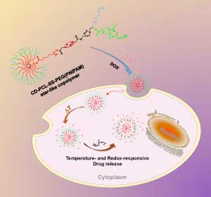
Nanoscale drug delivery particles that can sense their surroundings(Nanowerk News) A*STAR researchers have developed nanoscale drug delivery particles that can sense their surroundings, and release their payload only after entering a cell, a discovery that could make many existing medicines more effective (Polymer Chemistry, „“Y”-shape armed amphiphilic star-like copolymers: design, synthesis and dual-responsive unimolecular micelle formation for controlled drug delivery”). |
|
| The new nanoparticles, developed by Zibiao Li from the A*STAR Institute of Materials Research and Engineering and his collaborators, are a significant upgrade from previous generations of polymer-based drug delivery nanoparticles. Early examples typically consisted of simple polymer chains with a polar, hydrophilic head and a non-polar, hydrophobic tail. In water, these chains naturally aggregate into spheres, with their hydrophobic tails all pointing inward to form a non-polar core. The core formed a good site for drug molecules to nestle. In the bloodstream, however, these aggregates tend to be torn apart. | |
 |
|
| The change in temperature and chemical environment inside the cell triggers the nanoparticle to release its drug payload. (© RSC) | |
| Li and his colleagues used the latest polymer synthesis techniques to create single-molecule nanoparticles. Rather than a self-assembled aggregate of separate polymers, the team synthesized a more robust structure in which the polymer chains were strongly covalently bonded to a central core. The synthesis began with beta-cyclodextrin, a circular sugar molecule with 21 hydroxyl groups on its surface. The hydroxyl groups formed the chemical anchors from which the team constructed the nanoparticle’s multiple long, Y-shaped, multifunctional polymer arms. | |
| “The greatest challenge in making the [nanoparticle] was to integrate different synthetic methodologies into one macromolecular design,” says Li. At one of the ends of each Y-shaped branch, the team attached a temperature-sensitive polymer called PNIPAM. At room temperature the PNIPAM polymer extends outward, but collapses once body temperature, 37 degrees Celsius, is reached, allowing the nanoparticle’s drug molecule cargo to escape. | |
| Mid-way along each polymer arm, the team installed a sulfur-sulfur bond. When the particles enter a cell, they meet high levels of glutathione, a molecule that cleaves sulfur-sulfur bonds. Thus the nanoparticle’s outer polymer coat is snipped off, and the drug is released even faster. | |
| When the team tested this effect with an anti-cancer drug called doxorubicin, the dual-action effect was clear. “The change from room temperature to body temperature, and the effect of glutathione, showed synergistic and fast release of the drug,” Li says. At lower temperatures, or in the absence of glutathione, drug release was significantly slower, he says. | |
| “The next plan is to integrate new functionality, for precision release of the drug for targeted cancer therapy,” says Li. One end of each Y-shaped branch could be functionalized with a molecule that sticks selectively to cancer cells, delivering the drug precisely where it is needed. |
| Source: A*STAR |
Aceste cipuri ar putea fi, de asemenea, utilizate pentru a evalua medicamentele de anticorpi și alte imunoterapii, care sunt dificil de testat temeinic la animale deoarece sunt proiectate să interacționeze cu sistemul imunitar uman