Materialul cu auto-vindecare se poate construi din carbon în aer(Self-healing material can build itself from carbon in the air)

Un material proiectat de inginerii chimici ai MIT poate reacționa cu dioxidul de carbon din aer, să crească, să se consolideze și chiar să se repare

Self Healing material

Senzorii sensibili la nanofire oferă piele ultra-sensibilă pentru roboți(Flexible nanowire sensors give robots ultra-sensitive skin)

Universitatea din Texas de la Arlington a brevetat o piele inteligenta, creata de un cercetator UTA, care va da robotilor senzatii tactile mai sensibile decat oameni.Pielea inteligenta este de fapt alcatuita din milioane de senzori de nanofire flexibile care au mult mai multe informatii decat pielea oamenilor. Pe măsură ce senzorii perie o suprafață, robotul colectează toate informațiile pe care senzorii le trimit înapoi.Alte aplicații posibile viitoare includ aderarea pielii inteligente la proteze pentru a le dota cu un sentiment, aplicarea tehnologiei pe alte dispozitive medicale, țeserea pielii în uniforma unui soldat de luptă, astfel încât să poată fi detectată orice substanță chimică toxică sau identificarea amprentelor digitale.

Posted: Aug 09, 2018

Flexible nanowire sensors give robots ultra-sensitive skin

(Nanowerk News) The University of Texas at Arlington has patented a smart skin, created by a UTA researcher, that will give robots more sensitive tactile feeling than humans.

“The idea is to have robots work better alongside people,” said Zeynep Çelik-Butler, a UTA electrical engineering professor. “The smart skin is actually made up of millions of flexible nanowire sensors that take in so much more information than people’s skin. As the sensors brush against a surface, the robot collects all the information those sensors send back.”
Çelik-Butler said the sensors, which are flexible and made of zinc oxide nanorods, are self-powered and do not need any external voltage for operation. Each is about 0.2 microns in diameter, while a human hair is about 40 to 50 microns.
In addition, the developed sensors were fully packaged in a chemical and moisture resistant polyimide that greatly enhances usability in harsh environments. The result is a thin, flexible, self-powered tactile sensing layer, suitable as a robotic or prosthetic skin.
Smart skin
Smart skin technology. (Image: UTA)
The smart skin technology allows the robots to sense temperature changes and surface variations, which would allow a person alongside the robot to be safer or react accordingly.
Other possible future applications include adhering the smart skin to prosthetics to equip them with some feeling, applying the technology to other medical devices, weaving the skin into the uniform of a combat soldier so that any toxic chemicals could be detected or fingerprint identification.
“These sensors are highly sensitive and if they were brushed over a partial fingerprint, the technology could help identify who that person is,” Çelik-Butler said. “Imagine people being able to ascertain a person’s identity with this hairy robot, as my students call it.”
Teri Schultz, director of technology management in the UTA Office of Research, said the technology shows promise in a number of commercial sectors.
“Robots are the here and now,” Schultz said. “We could see this technology develop with the next generation of robots to allow them to be more productive in helping people.”
The research yielded an academic paper in a 2015 issue of IEEE Sensors Journal („Self-Powered Tactile Pressure Sensors Using Ordered Crystalline ZnO Nanorods on Flexible Substrates Toward Robotic Skin and Garments”). Funding for the smart skin technology has come from the National Science Foundation and NASA.
Source: University of Texas at Arlington

Oamenii de știință dezvoltă materiale unice pentru a repara organele și țesuturile deteriorate(Scientists develop unique materials to repair damaged organs and tissue)

a creat materiale polimerice unice pentru scopuri medicale care repară organe umane traumatizate ( Cell and Tissue Biology , Bioresorption of Porous 3D Matrice pe bază de colagen în ficat și țesut muscular ” ).
Specialiștii de laborator au dezvoltat un material poros tridimensional din colagen și chitosan, un analog al țesutului osos. Folosind acest material, cercetatorii pot restaura parti ale oaselor pierdute ca urmare a unor traume sau boli.

Posted: Aug 06, 2018

Scientists develop unique materials to repair damaged organs and tissue

(Nanowerk News) Tissue engineering is the future of medicine. Under Project 5-100, the Polymer Materials for Tissue Engineering and Transplantology Laboratory of Peter the Great St. Petersburg Polytechnic University (SPbPU) created unique polymeric materials for medical purposes that repair traumatized human organs (Cell and Tissue Biology, „Bioresorption of Porous 3D Matrices Based on Collagen in Liver and Muscular Tissue”).

The laboratory specialists developed a three-dimensional porous material made of collagen and chitosan, an analog of bone tissue. Using this material, the researchers can restore parts of bone lost as a result of trauma or illness.
According to the scientists, this is a completely new medical area not only in Russia but in the whole world. The terminology has not yet been defined. They are currently called „mimicking” materials, as they trick the body. The polymer matrix is implanted into damaged liver tissue, bones or vessels, saturated with the cells of these organs. Since the materials are made from biocompatible components (chitosan and collagen), the body is tricked and does not reject the foreign object. Over time, the matrix decomposes and the artificial tissue is replaced by natural tissue.
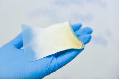
Biocompatible and Bioresorbable Polymer Materials
Scientists created biocompatible and bioresorbable polymer materials. (Image: Peter the Great St. Petersburg Polytechnic University)
„We are not deceiving nature, we are just helping it to cope with a medical problem. Experts are currently debating whether it is better to use an implant or restore an organ. A person with an artificial organ must take medication for the rest of their lifetime to prevent the body from rejecting it. This is not the case for tissue grown from human cells,” explains Vladimir Yudin, Head of the Laboratory.
The development of artificial organs for transplantology is an urgent priority of modern medicine. The successful development of this area largely depends on the creation of biocompatible and bioresorbable polymer materials.
The scientists of St. Petersburg Polytechnic University have not only developed the technology to create biocompatible materials that stimulate the restoration of natural tissues, but also managed to regulate the resorption time of the materials. It is very important for the implanted materials not to disintegrate before the new fabric is formed.
The results of preclinical studies showed that after a certain period of time, a three-dimensional sponge embedded in a bone starts to become covered with natural bone tissue, while the material itself decomposes. In addition, the developed collagen sponge was studied both in liver tissues and in muscle tissue – the material also stimulated the restoration of the natural tissue of the organs.
The researchers also developed wound covers, prostheses of blood vessels, and suture threads. In vivo preclinical trials were conducted with these materials, and the results proved that they are effective. The materials are recommended for use in tissue engineering and cellular transplantation.
Source: Peter the Great Saint-Petersburg Polytechnic University

Inginerii proiectează un bandaj de compresie care schimbă culoarea(Engineers design color-changing compression bandage)

Apoi, astfel de fibre ar putea fi filetate în bandaje, împreună cu textile, cum ar fi îmbrăcămintea atletică și pantofii ca indicatori de culoare pentru, de exemplu, tulpina musculară în timpul antrenamentelor. Kolle prevede că acestea pot fi, de asemenea, utilizate ca senzori de tensiune pentru infrastructură și mașini.

Posted: May 29, 2018

Engineers design color-changing compression bandage

(Nanowerk News) Compression therapy is a standard form of treatment for patients who suffer from venous ulcers and other conditions in which veins struggle to return blood from the lower extremities. Compression stockings and bandages, wrapped tightly around the affected limb, can help to stimulate blood flow. But there is currently no clear way to gauge whether a bandage is applying an optimal pressure for a given condition.

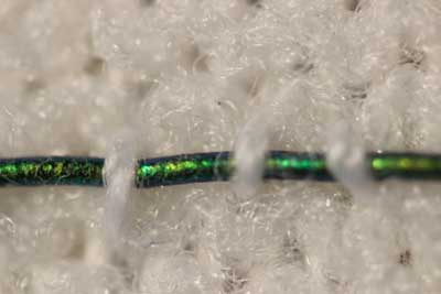
Now engineers at MIT have developed pressure-sensing photonic fibers that they have woven into a typical compression bandage. As the bandage is stretched, the fibers change color. Using a color chart, a caregiver can stretch a bandage until it matches the color for a desired pressure, before, say, wrapping it around a patient’s leg.
The photonic fibers can then serve as a continuous pressure sensor — if their color changes, caregivers or patients can use the color chart to determine whether and to what degree the bandage needs loosening or tightening.
“Getting the pressure right is critical in treating many medical conditions including venous ulcers, which affect several hundred thousand patients in the U.S. each year,” says Mathias Kolle, assistant professor of mechanical engineering at MIT. “These fibers can provide information about the pressure that the bandage exerts. We can design them so that for a specific desired pressure, the fibers reflect an easily distinguished color.”
pressure-sensing photonic fibers woven into a typical compression bandage
Engineers at MIT have developed pressure-sensing photonic fibers that they have woven into a typical compression bandage. (Courtesy of the researchers)
Kolle and his colleagues have published their results in the journal Advanced Healthcare Materials(„Stretchable Optomechanical Fiber Sensors for Pressure Determination in Compressive Medical Textiles”). Co-authors from MIT include first author Joseph Sandt, Marie Moudio, and Christian Argenti, along with J. Kenji Clark of the Univeristy of Tokyo, James Hardin of the United States Air Force Research Laboratory, Matthew Carty of Brigham and Women’s Hospital-Harvard Medical School, and Jennifer Lewis of Harvard University.

Natural inspiration

The color of the photonic fibers arises not from any intrinsic pigmentation, but from their carefully designed structural configuration. Each fiber is about 10 times the diameter of a human hair. The researchers fabricated the fiber from ultrathin layers of transparent rubber materials, which they rolled up to create a jelly-roll-type structure. Each layer within the roll is only a few hundred nanometers thick.
In this rolled-up configuration, light reflects off each interface between individual layers. With enough layers of consistent thickness, these reflections interact to strengthen some colors in the visible spectrum, for instance red, while diminishing the brightness of other colors. This makes the fiber appear a certain color, depending on the thickness of the layers within the fiber.
“Structural color is really neat, because you can get brighter, stronger colors than with inks or dyes just by using particular arrangements of transparent materials,” Sandt says. “These colors persist as long as the structure is maintained.”
The fibers’ design relies upon an optical phenomenon known as “interference,” in which light, reflected from a periodic stack of thin, transparent layers, can produce vibrant colors that depend on the stack’s geometric parameters and material composition. Optical interference is what produces colorful swirls in oily puddles and soap bubbles. It’s also what gives peacocks and butterflies their dazzling, shifting shades, as their feathers and wings are made from similarly periodic structures.
“My interest has always been in taking interesting structural elements that lie at the origin of nature’s most dazzling light manipulation strategies, to try recreating and employing them in useful applications,” Kolle says.

A multilayered approach

The team’s approach combines known optical design concepts with soft materials, to create dynamic photonic materials.
While a postdoc at Harvard in the group of Professor Joanna Aizenberg, Kolle was inspired by the work of Pete Vukusic, professor of biophotonics at the University of Exeter in the U.K., on Margaritaria nobilis, a tropical plant that produces extremely shiny blue berries. The fruits’ skin is made up of cells with a periodic cellulose structure, through which light can reflect to give the fruit its signature metallic blue color.
Together, Kolle and Vukusic sought ways to translate the fruit’s photonic architecture into a useful synthetic material. Ultimately, they fashioned multilayered fibers from stretchable materials, and assumed that stretching the fibers would change the individual layers’ thicknesses, enabling them to tune the fibers’ color. The results of these first efforts were published in Advanced Materials in 2013.
When Kolle joined the MIT faculty in the same year, he and his group, including Sandt, improved on the photonic fiber’s design and fabrication. In their current form, the fibers are made from layers of commonly used and widely available transparent rubbers, wrapped around highly stretchable fiber cores. Sandt fabricated each layer using spin-coating, a technique in which a rubber, dissolved into solution, is poured onto a spinning wheel. Excess material is flung off the wheel, leaving a thin, uniform coating, the thickness of which can be determined by the wheel’s speed.
For fiber fabrication, Sandt formed these two layers on top of a water-soluble film on a silicon wafer. He then submerged the wafer, with all three layers, in water to dissolve the water-soluble layer, leaving the two rubbery layers floating on the water’s surface. Finally, he carefully rolled the two transparent layers around a black rubber fiber, to produce the final colorful photonic fiber.

Reflecting pressure

The team can tune the thickness of the fibers’ layers to produce any desired color tuning, using standard optical modeling approaches customized for their fiber design.
“If you want a fiber to go from yellow to green, or blue, we can say, ‘This is how we have to lay out the fiber to give us this kind of [color] trajectory,’” Kolle says. “This is powerful because you might want to have something that reflects red to show a dangerously high strain, or green for ‘ok.’ We have that capacity.”
The team fabricated color-changing fibers with a tailored, strain-dependent color variation using the theoretical model, and then stitched them along the length of a conventional compression bandage, which they previously characterized to determine the pressure that the bandage generates when it’s stretched by a certain amount.
The team used the relationship between bandage stretch and pressure, and the correlation between fiber color and strain, to draw up a color chart, matching a fiber’s color (produced by a certain amount of stretching) to the pressure that is generated by the bandage.
To test the bandage’s effectiveness, Sandt and Moudio enlisted over a dozen student volunteers, who worked in pairs to apply three different compression bandages to each other’s legs: a plain bandage, a bandage threaded with photonic fibers, and a commercially-available bandage printed with rectangular patterns. This bandage is designed so that when it is applying an optimal pressure, users should see that the rectangles become squares.
Overall, the bandage woven with photonic fibers gave the clearest pressure feedback. Students were able to interpret the color of the fibers, and based on the color chart, apply a corresponding optimal pressure more accurately than either of the other bandages.
The researchers are now looking for ways to scale up the fiber fabrication process. Currently, they are able to make fibers that are several inches long. Ideally, they would like to produce meters or even kilometers of such fibers at a time.
“Currently, the fibers are costly, mostly because of the labor that goes into making them,” Kolle says. “The materials themselves are not worth much. If we could reel out kilometers of these fibers with relatively little work, then they would be dirt cheap.”
Then, such fibers could be threaded into bandages, along with textiles such as athletic apparel and shoes as color indicators for, say, muscle strain during workouts. Kolle envisions that they may also be used as remotely readable strain gauges for infrastructure and machinery.
“Of course, they could also be a scientific tool that could be used in a broader context, which we want to explore,” Kolle says.
Source: By Jennifer Chu, MIT

Mătase fluorescentă de culoare roșie poate ucide bacteriile dăunătoare ca remediu biologic și ecologic(Far-red fluorescent silk can kill harmful bacteria as biomedical and environmental remedy)

Un material hibrid de mătase atacă bacteriile atunci când este iluminat de o lumină verde, datorită cercetătorilor de proteină fluorescenți de departe roșii transferați la machiajul lor genetic.Materialul complet natural ar fi mai sigur decât mijloacele convenționale fotocatalitic sau activat cu lumină pentru a ucide agenții patogeni daunatori, cum ar fi bacteriile, care utilizează semiconductori potențial biohazard și necesită lumină ultravioletă care provoacă cancer pentru activare.Tehnologia de procesare a mătăsii fluorescente (mKate2) poate fi aplicată dezvoltării de bandaje inteligente pentru vindecarea rănilor pe pielea umană
O alternativă de mătase proiectată de Universitatea Purdue și Institutul Național de Cercetări Agricole coreene ar folosi biomateriale plasmonice asemănătoare biomaterialelor și lumină vizibilă, care, de asemenea, ajută la vindecarea rănilor și la reabilitarea mediului, inclusiv purificarea aerului și a apei.

Posted: Apr 19, 2018

Far-red fluorescent silk can kill harmful bacteria as biomedical and environmental remedy

(Nanowerk News) A silk hybrid material attacks bacteria when illuminated by a green light, thanks to a far-red fluorescent protein researchers transferred to its genetic makeup.

The all-natural material would be safer than conventional photocatalytic, or light-activated, means to kill harmful pathogens such as bacteria, which use potentially biohazardous semiconductors and require cancer-causing ultraviolet light for activation.
A silk alternative engineered by Purdue University and the Korean National Institute of Agricultural Research would instead use plasmonic photocatalyst-like biomaterials and visible light, which also aid in wound healing and environmental remediation including air and water purification. Their findings published in Advanced Science („Green-light-activated photoreaction via genetic hybridization of far-red fluorescent protein and silk”).
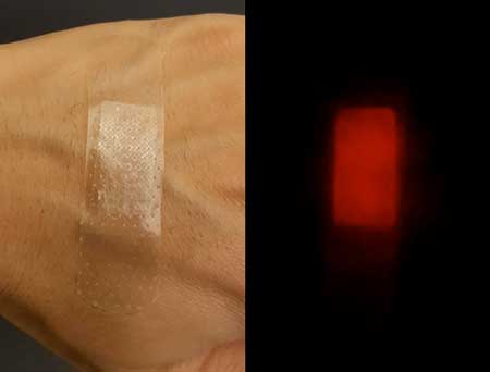
Silk Hybrid Bandage
The processing technology of fluorescent (mKate2) silk can be applied to developing smart medical bandages for wound healing on the human skin. (Image: Jung Woo Leem, Purdue University)
„Silk is an ancient and well-known biomaterial,” said Young Kim, Purdue associate professor of biomedical engineering. „It doesn’t have any issues with the human body. And the nice thing about green light is that it’s not harmful – the color corresponds to the strongest intensity of the solar spectrum.”
To combine the benefits of silk and green light, researchers inserted the gene for „mKate2,” a far-red fluorescent protein, into a silk host. Shining a green light on the resulting hybrid generates reactive oxygen species (ROS), which are effective radicals for breaking down organic contaminants and attacking the membrane and DNA of pathogens.
When E. coli on the fluorescent silk were illuminated by a weak green light for 60 minutes, the bacteria’s survival rate dropped to 45 percent.
The researchers found that the hybrid could be processed into a solution, film, bandage and fabric. „We’ve basically added fluorescence to silk to facilitate disinfection or decontamination using just visible light,” said Jung Woo Leem, a visiting scholar in Purdue’s school of biomedical engineering.
Kim’s team believes that green-light activated red fluorescent silk could be both more efficient and scalable than other plasmonic photocatalysts, in which metal nanoparticles hybridized from semiconductor materials also use visible light but could still pose negative environmental consequences.
„The silk photocatalysts would be easier and safer to produce than plasmonic ones since silkworms, rather than industrial facilities, provide the host for ROS-generating materials. It’s a completely new green manufacturing of nanomaterials,” Kim said.
Because ambient white light also includes green light, the researchers anticipate that the silk hybrid material should typically have a strong enough light source to generate ROS as long as a green light controls ROS generation.
Kim’s team plans to take advantage of silk’s biocompatibility with the human body both inside and out. „We’re thinking about some implantable and injectable wound healing materials that dissolve over time in the body. Then we wouldn’t need to do additional surgery to take it out,” Kim said.
Source: Purdue University

Cheie de mătase din Spider pentru un compozit nou de fixare a oaselor(Spider silk key to new bone-fixing composite)

au creat un compozit biodegradabil fabricat din fibre de mătase care pot fi utilizate pentru a repara oasele rupte încărcate fără complicații uneori prezentate de alte materiale.Echipa a început deja să testeze noi derivați ai compozitului, inclusiv acelea care încorporează o singură formă cristalină a hidroxiapatitei pentru o rezistență mai mare și o variație a amestecului de acoperire pentru a maximiza proprietățile sale mecanice pentru oasele care poartă o greutate mai mare.

Posted: Apr 19, 2018

Spider silk key to new bone-fixing composite

(Nanowerk News) University of Connecticut researchers have created a biodegradable composite made of silk fibers that can be used to repair broken load-bearing bones without the complications sometimes presented by other materials.

Repairing major load-bearing bones such as those in the leg can be a long and uncomfortable process.
To facilitate repair, doctors may install a metal plate to support the bone as it fuses and heals. Yet that can be problematic. Some metals leach ions into surrounding tissue, causing inflammation and irritation. Metals are also very stiff. If a metal plate bears too much load in the leg, the new bone may grow back weaker and be vulnerable to fracture.
Seeking a solution to the problem, UConn professor Mei Wei, a materials scientist and biomedical engineer, turned to spiders and moths for inspiration. In particular, Wei focused on silk fibroin, a protein found in the silk fibers spun by spiders and moths known for its toughness and tensile strength.
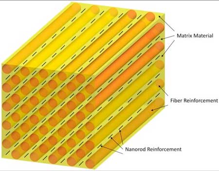
Biodegradable Bone-Fixing Composite Made Partly of Silk Fibers
A 3-D rendering of a novel bone-fixing composite developed at the University of Connecticut. The composite is made of silk fibers and fibers of polylactic acid that are coated with fine bioceramic particles to add strength while maintaining flexibility. The biodegradable composite helps heal bones without the complications posed by metal parts. (Image: Bryant Heimbach/UConn)
The medical community has been aware of silk fibroin for a while. It is a common component in medical sutures and tissue engineering because of its strength and biodegradability. Yet no one had ever tried to make a dense polymer composite out of it, and that is what Wei knew she needed if she was going to create a better device for healing broken load-bearing bones.
Working with UConn associate professor Dianyun Zhang, a mechanical engineer, Wei’s lab began testing silk fibroin in various composite forms, looking for the right combination and proportion of different materials to achieve optimum strength and flexibility. The new composite certainly needed to be strong and stiff, yet not so much so that it would inhibit dense bone growth. At the same time, the composite needed to be flexible, allowing patients to retain their natural range of motion and mobility while the bone healed.
After dozens of tests, Wei and Zhang found the materials they were looking for. The new composite consists of long silk fibers and fibers of polylactic acid – a biodegradable thermoplastic derived from cornstarch and sugar cane – that are dipped in a solution in which each is coated with fine bioceramic particles made of hydroxyapatite (the calcium phosphate mineral found in teeth and bones). The coated fibers are then packed in layers on a small steel frame and pressed into a dense composite bar in a hot compression mold.
In a study recently published in the Journal of the Mechanical Behavior of Biomedical Materials(„High performance resorbable composites for load-bearing bone fixation devices”), Wei reports that the high-performance biodegradable composite showed strength and flexibility characteristics that are among the highest ever recorded for similar bioresorbable materials in literature.
And they could get even better.
„Our results are really high in terms of strength and flexibility, but we feel that if we can get every component to do what we want them to do, we can get even higher,” says Wei, who also serves as the School of Engineering’s associate dean for research and graduate education.
The new composite is also resilient. Large leg bones in adults and seniors can take many months to heal. The composite developed in Wei’s lab does its job and then starts to degrade after a year. No surgery is required for removal.
Joining Wei and Zhang in the research were Bryant Heimbach, a Ph.D. candidate and materials scientist in Wei’s lab; and Beril Tonyali, a UConn undergraduate pursuing a degree in materials science and engineering.
The team has already begun testing new derivatives of the composite, including those that incorporate a single crystalline form of the hydroxyapatite for greater strength and a variation of the coating mixture to maximize its mechanical properties for bones bearing more weight.
Source: University of Connecticut

Spikele de grafen se pot ucide pe bacterii pe implanturi(Spikes of graphene can kill bacteria on implants)

Un strat mic de fulgi de grafen se transforma intr-o arma mortala si ucide bacteriile, oprindu-se infectiile in timpul unor proceduri precum chirurgia implantuluiO echipă de cercetare de la Chalmers a arătat acum că un strat de fulgi de grafeni verticali formează o suprafață protectoare care face imposibilă atașarea bacteriilor. În schimb, bacteriile sunt despicate în afară de fulgi ascuți de grafen și uciși. Acoperirea implanturilor cu un strat de fulgi grafenici poate, prin urmare, ajuta la protejarea pacientului împotriva infecțiilor, la eliminarea necesității tratamentului antibiotic și la reducerea riscului de respingere a implantului.

Posted: Apr 16, 2018

Spikes of graphene can kill bacteria on implants

(Nanowerk News) A tiny layer of graphene flakes becomes a deadly weapon and kills bacteria, stopping infections during procedures such as implant surgery. This is the findings of new research from Chalmers University of Technology, Sweden, recently published in the scientific journal Advanced Materials Interfaces („Vertically Aligned Graphene Coating is Bactericidal and Prevents the Formation of Bacterial Biofilms”).

Operations for surgical implants, such as hip and knee replacements or dental implants, have increased in recent years. However, in such procedures, there is always a risk of bacterial infection. In the worst case scenario, this can cause the implant to not attach to the skeleton, meaning it must be removed.
Bacteria travel around in fluids, such as blood, looking for a surface to cling on to. Once in place, they start to grow and propagate, forming a protective layer, known as a biofilm.
A research team at Chalmers has now shown that a layer of vertical graphene flakes forms a protective surface that makes it impossible for bacteria to attach. Instead, bacteria are sliced apart by the sharp graphene flakes and killed. Coating implants with a layer of graphene flakes can therefore help protect the patient against infection, eliminate the need for antibiotic treatment, and reduce the risk of implant rejection. The osseointegration – the process by which the bone structure grow to attach the implant – is not disturbed. In fact, the graphene has been shown to benefit the bone cells.
Spikes of Graphene Slice Bacteria Apart, but Leave Larger Human Cells Undamaged
Vertical graphene flakes form a protective surface that makes it impossible for bacteria to attach. Instead, bacteria are sliced apart by the sharp graphene flakes and killed. Human cells volume is typically 15,000 times larger. So, what constitutes a deadly knife attack for a bacterium, is therefore only a tiny scratch for a human cell. Coating implants with a layer of graphene flakes can therefore help protect the patient against infection, eliminate the need for antibiotic treatment, and reduce the risk of implant rejection. The osseointegration – the process by which the bone structure grows to attach the implant – is not disturbed. In fact, the graphene has been shown to benefit the bone cells. (Image: Yen Strandqvist/Chalmers University of Technology)
Chalmers University is a leader in the area of graphene research, but the biological applications did not begin to materialise until a few years ago. The researchers saw conflicting results in earlier studies. Some showed that graphene damaged the bacteria, others that they were not affected.
„We discovered that the key parameter is to orient the graphene vertically. If it is horizontal, the bacteria are not harmed” says Ivan Mijakovic, Professor at the Department of Biology and Biological Engineering.
The sharp flakes do not damage human cells. The reason is simple: one bacterium is one micrometer – one thousandth of a millimeter – in diameter, while a human cell is 25 micrometers. So, what constitutes a deadly knife attack for a bacterium, is therefore only a tiny scratch for a human cell.
„Graphene has high potential for health applications. But more research is needed before we can claim it is entirely safe. Among other things, we know that graphene does not degrade easily” says Jie Sun, Associate Professor at the Department of Micro Technology and Nanoscience.
Good bacteria are also killed by the graphene. But that’s not a problem, as the effect is localised and the balance of microflora in the body remains undisturbed.
„We want to prevent bacteria from creating an infection. Otherwise, you may need antibiotics, which could disrupt the balance of normal bacteria and also enhance the risk of antimicrobial resistance by pathogens” says Santosh Pandit, postdoc at Biology and Biological Engineering.
Vertical flakes of graphene are not a new invention, having existed for a few years. But the Chalmers research teams are the first to use the vertical graphene in this way. The next step for the research team will be to test the graphene flakes further, by coating implant surfaces and studying the effect on animal cells.
Chalmers cooperated with Wellspect Healthcare, a company which makes catheters and other medical instruments, in this research. They will now continue with a second study. The projects are funded by Vinnova (a Swedish government agency).

The making of vertical graphene

Graphene is made of carbon atoms. It is only a single atomic layer thick, and therefore the world’s thinnest material. Graphene is made in flakes or films. It is 200 times stronger than steel and has very good conductivity thanks to its rapid electron mobility. Graphene is also extremely sensitive to molecules, which allows it to be used in sensors.
Graphene can be made by CVD, or Chemical Vapor Deposition. The method is used to create a thin surface coating on a sample. The sample is placed in a vacuum chamber and heated to a high temperature at the same time as three gases – usually hydrogen, methane and argon – are released into the chamber. The high heat causes gas molecules to react with each other, and a thin layer of carbon atoms is created.
To produce vertical graphene forms, a process known as Plasma-Enhanced Chemical Vapor Deposition, or PECVD, is used. Then, an electric field – a plasma – is applied over the sample, which causes the gas to be ionized near the surface. With the plasma, the layer of carbon grows vertically from the surface, instead of horizontally as with CVD.
Source: Chalmers University of Technology

Pansamentele din nanofibre vindecă rănile, promovează regenerarea(Nanofiber dressings heal wounds, promote regeneration)

au dezvoltat pansamente noi rana, care accelereaza dramatic vindecarea si imbunatatirea regenerarii tesutului. Cele două tipuri diferite de pansamente nanofibre, descrise în lucrări separate, utilizează proteine ​​care apar în plante și animale pentru a promova vindecarea și regenerarea țesuturilor.
„Sistemul nostru de fabricație a fibrelor a fost dezvoltat special pentru a dezvolta terapeutica pentru rănile de război”Aceste descoperiri arata marea promisiune a nanofibrelor pe baza de soia pentru vindecarea ranilor,Ambele tipuri de îmbrăcăminte, potrivit cercetătorilor, au avantaje în spațiul de vindecare a rănilor. Nanofibrele pe bază de soia – constând din acetat de celuloză și hidrolizat de proteine ​​din soia – sunt ieftine, făcându-le o opțiune bună pentru utilizarea pe scară largă, cum ar fi arsurile. Pansamentele de fibronectină, pe de altă parte, ar putea fi folosite pentru răni mai mici pe față și pe mâini, unde prevenirea cicatrizării este importantă.

Posted: Mar 19, 2018

Nanofiber dressings heal wounds, promote regeneration

(Nanowerk News) Researchers from the Harvard John A. Paulson School of Engineering and Applied Sciences (SEAS) and the Wyss Institute for Biologically Inspired Engineering have developed new wound dressings that dramatically accelerate healing and improve tissue regeneration. The two different types of nanofiber dressings, described in separate papers, use naturally-occurring proteins in plants and animals to promote healing and regrow tissue.

„Our fiber manufacturing system was developed specifically for the purpose of developing therapeutics for the wounds of war,” said Kit Parker, the Tarr Family Professor of Bioengineering and Applied Physics at SEAS and senior author of the research. „As a soldier in Afghanistan, I witnessed horrible wounds and, at times, the healing process for those wounds was a horror unto itself. This research is a years-long effort by many people on my team to help with these problems.”
Parker is also a Core Faculty Member of the Wyss Institute.
The most recent paper, published in Biomaterials („Production-scale fibronectin nanofibers promote wound closure and tissue repair in a dermal mouse model”), describes a wound dressing inspired by fetal tissue.
Hair Follicle Regeneration
This is a tissue section 14 days post-wounding reveals the presence of hair follicles regenerating at the center of the wound. Re-growing hair follicles is one of the biggest challenges in the field of wound healing. (Image: Disease Biophysics Group/Harvard University)
In the late 1970s, when scientists first started studying the wound healing process early in development, they discovered something unexpected: wounds incurred before the third trimester left no scars. This opened a range of possibilities for regenerative medicine. But for decades, researchers have struggled to replicate those unique properties of fetal skin.
Unlike adult skin, fetal skin has high levels of a protein called fibronectin, which assembles into the extracellular matrix and promotes cell binding and adhesion. Fibronectin has two structures: globular, which is found in blood, and fibrous, which is found in tissue. Even though fibrous fibronectin holds the most promise for wound healing, previous research focused on the globular structure, in part because manufacturing fibrous fibronectin was a major engineering challenge.
But Parker and his team are pioneers in the field of nanofiber engineering.
The researchers made fibrous fibronectin using a fiber manufacturing platform called Rotary Jet-Spinning (RJS), developed by Parker’s Disease Biophysics Group. RJS works likes a cotton candy machine – a liquid polymer solution, in this case globular fibronectin dissolved in a solvent, is loaded into a reservoir and pushed out through a tiny opening by centrifugal force as the device spins. As the solution leaves the reservoir, the solvent evaporates and the polymers solidify. The centrifugal force unfolds the globular protein into small, thin fibers. These fibers – less than one micrometer in diameter – can be collected to form a large-scale wound dressing or bandage.
„The dressing integrates into the wound and acts like an instructive scaffold, recruiting different stem cells that are relevant for regeneration and assisting in the healing process before being absorbed into the body,” said Christophe Chantre, a graduate student in the Disease Biophysics Group and first author of the paper.
In in vivo testing, the researchers found that wounds treated with the fibronectin dressing showed 84 percent tissue restoration within 20 days, compared to 55.6 percent restoration in wounds treated with a standard dressing.
The researchers also demonstrated that wounds treated with the fibronectin dressing have close to normal epidermal thickness and dermal architecture, and even regrew hair follicles – often considered one of the biggest challenges in the field of wound healing.
„This is an important step forward,” said Chantre. „Most work done on skin regeneration to date involves complex treatments combining scaffolds, cells and even growth factors. Here we were able to demonstrate tissue repair and hair follicle regeneration using an entirely material approach. This has clear advantages for clinical translation.”
In another paper published in Advanced Healthcare Materials („Soy Protein/Cellulose Nanofiber Scaffolds Mimicking Skin Extracellular Matrix for Enhanced Wound Healing”), the Disease Biophysics Group demonstrated a soy-based nanofiber that also enhances and promotes wound healing.
Soy protein contains both estrogen-like molecules – which have been shown to accelerate wound healing – and bioactive molecules similar to those that build and support human cells.
„Both the soy and fibronectin fiber technologies owe their success to keen observations in reproductive medicine,” said Parker. „During a woman’s cycle, when her estrogen levels go high, a cut will heal faster. If you do a surgery on a baby still in the womb, they have scar-less wound healing. Both of these new technologies are rooted in the most fascinating of all the topics in human biology – how we reproduce.”
In a similar way to fibronectin fibers, the research team used RJS to spin ultra-thin soy fibers into wound dressings. In experiments, the soy and cellulose-based dressing demonstrated a 72-percent increase in healing over wounds with no dressing and a 21-percent increase in healing over wounds dressed without soy protein.
„These findings show the great promise of soy-based nanofibers for wound healing,” said Seungkuk Ahn, a graduate student in the Disease Biophysics Group and first author of the paper. „These one-step, cost-effective scaffolds could be the next generation of regenerative dressings and push the envelope of nanofiber technology and the wound care market.”
Both kinds of dressing, according to researchers, have advantages in the wound-healing space. The soy-based nanofibers – consisting of cellulose acetate and soy protein hydrolysate – are inexpensive, making them a good option for large-scale usage, such as on burns. The fibronectin dressings, on the other hand, could be used for smaller wounds on the face and hands, where the prevention of scarring is important.
Source: Harvard John A. Paulson School of Engineering and Applied Sciences

Firele din nanotuburi de carbon intaresc nervii pentru tratamentele electrochirurgie si diagnosticare(Carbon nanotube yarn taps nerves for electroceutical treatments and diagnostics)

Acum, cercetările finanțate de NIBIB au transformat nanotuburile de carbon în fire sau fire flexibile, de natură nervoasă, capabile de legături de lungă durată de înaltă fidelitate la animalele vii. Dezvoltarea acestor fire biocompatibile deschide posibilitatea unor noi diagnostice și terapii bioelectrice prin reglarea funcției interne a organelor la nivelul nervului unic.
Nanotuburile individuale de carbon sunt trase de pe un substrat și se rotesc în firele flexibile de nanotuburi de carbon

Posted: Feb 26, 2018

Carbon nanotube yarn taps nerves for electroceutical treatments and diagnostics

(Nanowerk News) Ingested or injected pharmaceuticals can target specific molecules involved in disease processes, but get distributed throughout the body where they can cause unwanted side effects. An approach known as electroceuticals aims to avoid systemic exposure by using small wires to electrically monitor and manipulate individual nerves that control organ function and carry information about disease. Despite the promise of electroceuticals, it has been challenging to develop long-term therapies due to the lack of biocompatible wires.

Now, NIBIB-funded researches have spun carbon nanotubes into flexible, nerve-sized wires or yarns capable of high-fidelity long-term connections in live animals. The development of these biocompatible yarns opens the possibility of new bioelectric diagnostics and therapies through regulation of internal organ function at the single nerve level.

Individual carbon nanotubes are pulled from a substrate and spun into the flexible carbon nanotube yarn

Individual carbon nanotubes are pulled from a substrate and spun into the flexible carbon nanotube yarn. (© NPG)

All the organs of the body such as the heart, lungs, liver, and kidneys are automatically controlled by nerves that stretch from the brainstem to each organ. These nerves control organ functions such as heartbeat, breathing rates, and blood pressure, making constant adjustments in response to environmental and physiological changes. Variations in the electrical activity in this area of the brain, known as the autonomic nervous system, can also be predictors, indicators or causes of disease development.

“Monitoring and manipulating the autonomic nervous system to both understand and potentially treat disease has been an intriguing yet understudied approach to medicine,” explains Michael Wolfson, Ph.D., director of the NIBIB program in Rehabilitation Engineering and Implantable Medical Devices. “This is largely due to technical limitations in being able to insert wires into nerves that can reliably and safely record electrical activity over long periods of time and under different physiological conditions.”

In a study reported in the journal Scientific Reports („Chronic interfacing with the autonomic nervous system using carbon nanotube (CNT) yarn electrodes”), bioengineers at Case Western Reserve University in Cleveland, Ohio describe the development of highly flexible carbon nanotube (CNT) yarn electrodes that were capable of months-long electrical recording in major nerves of the autonomic nervous system in rats.

The CNT yarns are essentially, just that, yarns made from a “forest” of hundreds of carbon nanotubes that are pulled from the metal surface they are grown on and spun into a highly flexible, highly conductive wire 1/100th the size of a human hair.

carbon nanotube yarn is wound around a tungsten needle for insertion into a nerve

The carbon nanotube yarn is wound around a tungsten needle for insertion into a nerve. The coiling of the yarn causes it to remain solidly embedded in the nerve after the needle is withdrawn. Actual needle and yarn (top). Diagram showing detail of how yarn is wrapped around the needle for insertion (bottom). (© NPG)

Current technologies for recording electrical signals from nerves include relatively large stiff tungsten needles used by neurologists to obtain readings from single nerves of patients for several hours, but must be removed before causing lasting nerve damage. Other wire electrode technologies can record nerve signals for short periods but because of their dimensions and stiff mechanical properties they are not suitable for long-term recording in small nerves.

“The currently available electrode technologies simply do not match the mechanical properties of nerves,” explains Dominique Durand, Ph.D., Professor of Biomedical Engineering at Case Western and senior researcher on the CNT yarn work. “With those types of electrodes, it’s often like sticking glass into spaghetti. Our CNT yarns are similar in size and flexibility to actual nerves. These properties allow them to be stealthily inserted into specific nerves and remain there for months without destroying the tissue or inducing an attack by the immune system.”

The combination of the biocompatibility of the CNT yarns and their outstanding ability to carry an electrical signal that is 10 times stronger than current technologies makes them ideal for long term recording of specific nerve signals. Finally, the lack of nerve damage keeps the surrounding axons intact, which helps to eliminate background noise. Thus, the CNT yarns have an excellent signal to noise ratio (SNR), which is critical for this type of research.

The group tested the CNT yarns in two major nerves in the autonomic system in rats. One study involved the vagus nerve, which stretches throughout the body to connect to numerous organs. The nerve is known to control and monitor a range of functions including heart rate, digestive tract movement, sweating, and immune response.

CNT yarn electrodes were also inserted into the glossopharyngeal nerve. The nerve is connected to a number of organs including the carotid artery, and parts of the ear, tongue and salivary glands where it is known to be involved in swallowing.

Carbon nanotube yarn embedded into the vagus nerve of a rat

Carbon nanotube yarn embedded into the vagus nerve of a rat. (© NPG)

In both nerves, recordings of stable electrical activity were maintained over a 10-week period. Pulses of nerve activity were also monitored while the animal responded to physiological challenges. The challenges included distension of the rat’s stomach with saline solution, and short durations where the rats were in low oxygen environments. In each case, the physiological changes induced by the challenge resulted in easily detectable changes in electrical activity that were recorded using the CNT yarn implants over the entire 10-week period of the experiments.

“Although this is early research, we believe the results demonstrate that this technology can be used for reliable long term electrical monitoring of physiological functions,” says Durand. “This is an important step in pursuing monitoring of disease progression through the nerves that control the function of the diseased organ.”

Durand explains that the goal is to learn what electrical signatures or profiles are indicative of disease development and to use that for early intervention, potentially by electrical stimulation or even blocking of the nerve. For example, one therapy for severe hypertension is the cutting of the renal nerve. This is obviously irreversible. Durand, explained that CNT yarn electrodes could be used to block the nerve impulse without cutting the nerve, making the treatment reversible.

The group is also excited about the potential of this recording method to improve prosthetics. “When an arm has been amputated there are tens of thousands of neurons remaining,” said Durand. “The approach would be to insert the CNT yarns into individual nerves and record the electrical signals that are created as the individual thinks about moving the missing arm—essentially learning the electrical signals that are formed by the intention to move the arm.” The ultimate goal is to develop neural interfaces capable of translating those electrical signals into better control of the prosthetic by the user.

Source: National Institute of Biomedical Imaging and Bioengineering

Biocompatibile fibre de nanotehnologie anti-ardere care acționează ca „bandaje vii”(Biocompatible anti-burn nanotechnology fibers that act as ‘living bandages’)

Echipa de cercetători în domeniul nanotehnologiei a reușit să creeze „bandaje” din mai multe straturi fabricate din fibre biodegradabile și nanofilme bioactive multifuncționale, care [bandaje] previne cicatrizarea și accelerează regenerarea țesuturilor.Adăugarea efectului antibacterian prin introducerea de nanoparticule de argint sau prin îmbinarea antibioticelor, precum și creșterea activității biologice la suprafața grupelor hidrofilice (-COOH) și a proteinelor din plasmă sanguină au oferit materialului proprietăți unice de vindecare.
Sa observat o accelerare semnificativă a procesului de vindecare, regenerarea cu succes a țesutului normal acoperit cu piele și prevenirea cicatrizării pe pielea arsă sau deteriorată la aplicarea acestor bandajeri din materialul dezvoltat într-o zonă afectată

Posted: Feb 16, 2018

Biocompatible anti-burn nanotechnology fibers that act as ‘living bandages’

(Nanowerk News) In regenerative medicine, and particularly in burn therapy, the effective regeneration of damaged skin tissue and the prevention of scarring are usually the main goals. Scars form when skin is badly damaged, whether through a cut, burn, or a skin problem such as acne or fungal infection.

Scar tissue mainly consists of irreversible collagen and significantly differs from the tissue it replaces, having reduced functional properties. For example, scars on skin are more sensitive to ultraviolet radiation, are not elastic, and the sweat glands and hair follicles are not restored in the area.
The solution of this medical problem was proposed by the researchers from the NUST MISIS Inorganic Nanomaterials Laboratory, led by PhD Anton Manakhov, a senior researcher. The team of nanotechnology scientists has managed to create multi-layer ‘bandages’ made of biodegradable fibers and multifunctional bioactive nanofilms, which [the bandages] prevent scarring and accelerate tissue regeneration.
Multi-layer bandages made of biodegradable fibers and multifunctional bioactive nanofilms
Multi-layer bandages made of biodegradable fibers and multifunctional bioactive nanofilms. (Image: NUST MSIS)
The addition of the antibacterial effect by the introduction of silver nanoparticles or joining antibiotics, as well as the increase of biological activity to the surface of hydrophilic groups (-COOH) and the blood plasma proteins have provided unique healing properties to the material.
A significant acceleration of the healing process, the successful regeneration of normal skin covering tissue, and the prevention of scarring on the site of burnt or damaged skin have been observed when applying these bandages made of the developed material to an injured area. The antibacterial components of multifunctional nanofibers decrease inflammation, and the blood plasma with an increased platelet level — vital and multi-purposed for every element in the healing process — stimulates the regeneration of tissues. The bandages should not be removed or changed during treatment as it may cause additional pain to the patient. After a certain period of time, the biodegradable fiber simply “dissolves” without any side effects.
“With the help of chemical bonds, we were able to create a stable layer containing blood plasma components (growth factors, fibrinogens, and other important proteins that promote cell growth) on a polycaprolactone base. The base fibers were synthesized by electroforming. Then, with the help of plasma treatment, to increase the material`s hydrophilic properties, a polymer layer containing carboxyl groups was applied to the surface. The resulting layer was enriched with antibacterial and protein components”, noted Elizabeth Permyakova, one of the project members and laboratory scientists.
The research team has already conducted a series of pre-clinical trials jointly with the Research Institute of Experimental and Clinical Medicine (Novosibirsk, Russia). In vitro results have shown that with the application of these innovative bandages the regeneration process has been accelerated twice as quickly. In the near future, the team expects to get results of in vivo drug testing.
Source: NUST MSIS