Acoperire pe bază de oxid de grafen, inspirată de piele, care este la fel de dură ca și dinții și se poate vindeca(Skin-inspired graphene oxide based coating that’s as hard as teeth and can heal itself)

Mimetizând structura pielii umane, cercetătorii au folosit o tehnică strat-cu-strat pentru a forma un strat subțire dinamic, conținând alcool polivinilic și acid tanic. Stratul exterior dur conținea acești compuși plus un strat de oxid de grafen. Când este fabricat la o anumită grosime, materialul sa vindecat cu succes atunci când a fost tăiat și, de asemenea, ar putea ucide bacteriile.
Materialul ar putea într-o bună zi să servească ca o piele electronică sau chiar ca o acoperire rezistentă la zgârieturi pe clădiri sau pe telefoane.

Posted: Jan 31, 2018

Skin-inspired graphene oxide based coating that’s as hard as teeth and can heal itself

(Nanowerk News) Self-healing smart coatings could someday make scratches on cell phones a thing of the past. But researchers often have to compromise between strength and the ability to self-repair when developing these materials.

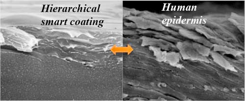
Now, one group reports in ACS Nano („An Epidermis-like Hierarchical Smart Coating with a Hardness of Tooth Enamel”) the development of a smart coating that is as hard as tooth enamel on the outside but can heal itself like skin can.
A quasi-linear layer-by-layer (LBL) film with constituent graphene oxide is deposited on top of an exponential LBL counterpart as a protective hard layer, forming a hierarchical stratified assembly mimicking the structure of epidermis
A quasi-linear layer-by-layer (LBL) film with constituent graphene oxide is deposited on top of an exponential LBL counterpart as a protective hard layer, forming a hierarchical stratified assembly mimicking the structure of epidermis. (© ACS)
The smart coating market is a booming industry and is only expected to grow in upcoming years. The most common smart coatings that can heal themselves are based on soft polymers that can wear out quickly. But hard coatings can be too rigid to come back together to fix a tear or scratch.
In previous research, Ming Yang and colleagues produced a stiffer, more healable coating, but its performance still needs to be optimized. In the current paper, the researchers developed a different way to make a soft, yet hard, self-healing material.
Mimicking the structure of human skin, the researchers used a layer-by-layer technique to form a soft, dynamic under layer containing polyvinyl alcohol and tannic acid. The hard outer layer contained these compounds plus a layer of graphene oxide. When fabricated at a certain thickness, the material successfully healed itself when cut, and it also could kill bacteria.
The material could someday serve as an electronic skin or even as a scratch-proof coating on buildings or phones.
Source: American Chemical Society

Masele musculare noi reacționează la lumina vizibilă(New molecular muscle responds to visible light)

au creat un tip complet nou de mușchi moleculară artificială dintr-un polimer capabil de ridicare greu;Cercetătorii se uită acum la modul în care își pot împerechea polimerul funcțional nou cu alții care sunt mai rezistenți și capabili să ridice sarcini mai grele. Ei vor, de asemenea, să dau seama cum să controleze mușchii artificiale moleculare folosind electrozi. Această acțiune ar fi similară cu modul în care semnalele electrice sunt transmise în organism și ar putea deschide calea spre viitoarele aplicații protetice.

Posted: Jan 29, 2018

New molecular muscle responds to visible light

(Nanowerk News) Picture a tiny, makeshift muscle that can curl a 20 milligram suspended weight when exposed to light. Under the right conditions, another mix packs enough power to bench-press a dime.

Researchers at Washington University in St. Louis have created a completely new kind of artificial molecular muscle from a polymer that’s capable of some heavy lifting — relatively speaking.
“The external trigger that initiates the actuation process can be something as simple as sunlight,” said Jonathan Barnes, assistant professor of chemistry in Arts & Sciences and a 2017 Packard Fellow. The novel polymer, which changes color and contracts when exposed to visible light, is described in a Jan. 24 publication of a special issue of Macromolecular Rapid Communications(„Photoredox-Based Actuation of an Artificial Molecular Muscle”).
Molecular Muscle Responds to Visible Light
The polymer was attached to a slip of black electrical tape. Exposed to blue light, the artificial molecular muscle moved a 20 milligram weight several centimeters from its starting position. (Image: Barnes laboratory, Washington University in St. Louis)
Barnes and his team have been working on their proof of concept for the novel redox-responsive polymer — one that contracts when electrons are added (reduction) and expands when they are taken away (oxidation) — since he started at Washington University less than two years ago.
Last fall, they demonstrated that they could successfully build their functional polymer and incorporate it into a pliant, bulk material called a hydrogel. The resulting material could be contracted to one-tenth its original volume and then expanded back to its original size, its long polymer chains delicately folding and unfolding in three dimensions.
The hydrogel contains 5 percent polymer overall, of which only 5 percent is the new, functional polymer; the rest is just water. This means that only 0.25 percent of the total hydrogel is the functional polymer, an incredibly low number in the field.
“If you look at other materials, the active polymer is usually in every link,” said Angelique Greene, a postdoctoral fellow in the Barnes laboratory. “Ours is very dilute, and yet our hydrogels still performed at a comparable and sometimes even better rate.”

Pulling their own weight

But the molecular muscle still needed to be triggered by chemical reduction in a wet solution. To address the slosh factor, the researchers then introduced visible-light-absorbing photoredox catalysts, embedded in the gel, and moved their muscles onto dry ground.
It was time for a strength test.
“We wanted to demonstrate that it could not only change shape, or bend, or turn a different color, but actually do work,” Barnes said.
The researchers affixed their best-performing gel to a piece of black electrical tape, and then attached a small, light piece of aluminum wire holding a small 20 milligram weight on the bottom. They exposed it to a blue light, and, after five hours, the polymer had moved the suspended weight several centimeters from its starting position.
“Here we have a lot fine control,” said Kevin Liles, a PhD candidate in chemistry who co-wrote the new study, along with Greene. “We can irradiate the polymer for a certain amount of time, stop it at a certain number of degrees (of bend), or irradiate a certain portion and get it to contract in certain areas.”
Five hours might seem like a long time to move a few centimeters, but Barnes isn’t worried that Mother Nature does it faster.
“If you’ve ever seen a flower or plant on the side of a mountain, it always bends toward where the light is,” Barnes said. “Nature finds a way to adapt to optimize the amount of light source that hits its petals. This material in principle does the exact same thing.”
The researchers are now looking at how to pair their novel functional polymer with others that are tougher and capable of lifting heavier loads. They also want to figure out how to control the artificial molecular muscles using electrodes. This action would be similar to the way that electrical signals are transmitted in the body, and could pave the way toward future prosthetic applications.
Source: Washington University in St. Louis

Un nanofenomen care declanșează procesul de reparare a oaselor(A nanophenomenon that triggers the bone-repair process)

Aceste rezultate au promisiunea pentru industria protezelor, în care noi materiale care reproduc sau amplifică acest efect flexoelectric pot fi folosite pentru a ghida regenerarea tisulară, conducând la o asimilare mai reușită a implanturilor.Prin studierea gradientilor de tulpina in oase si minerale osoase pure (hidroxiapatita), cercetatorii au fost in masura sa calculeze magnitudinea exacta a acestui camp electric. Descoperirile lor indică faptul că este suficient de mare în interiorul celor 50 microni necesari pentru vârful crackului, pentru a fi detectat de către celulele responsabile de repararea oaselor, implicând direct în acest proces flexoelectricitatea.

Posted: Jan 19, 2018

A nanophenomenon that triggers the bone-repair process

(Nanowerk News) Researchers at the Institut Català de Nanociència i Nanotecnologia (ICN2), a Severo Ochoa research centre located in the Universitat Autònoma de Barcelona (UAB) Campus and member of the Barcelona Institute of Science and Technology (BIST), have discovered that bone too is flexoelectric. They posit the possible role of flexoelectricity in the regeneration of bone tissue in and around the kind of microfractures incurred in bones on a daily basis.

The ICN2 Oxide Nanophysics Group led by ICREA Prof. Gustau Catalan reports these findings today in Advanced Materials („Flexoelectricity in Bones”), with lead author Fabián Vásquez-Sancho. The work has potential implications for the prosthetics industry and the development of biomimetic self-healing materials.
micro-cracks in bone mineral
Strain gradients can be large around small defects such as micro-cracks in bone mineral, so gradient-induced electricity (flexoelectricity) is also expected to be large around such defects.
Bones were already known to generate electricity under pressure, stimulating self-repair and remodelling. First reported in the late fifties, this was initially attributed to the piezoelectricity of bone’s organic component, collagen. However, studies have since observed markers of bone repair in the absence of collagen, suggesting that other effects are at play. In this work ICN2 researchers have revealed just such an effect: the flexoelectricity of bone’s mineral component.
Flexoelectricity is a property of some materials that causes them to emit a small voltage upon application of a non-uniform pressure. This response is extremely localised, becoming weaker as you move away from the point of maximum stress. In microfractures it is localised to the leading edge or tip of the crack, an atomically small site that, by definition, concentrates the maximum strain a material is able to withstand before full rupture. The result is a flexoelectric field of such magnitude that, in the immediate vicinity of the crack, it eclipses any background collagen piezoelectric effect.
By studying strain gradients in bones and pure bone mineral (hydroxyapatite), the researchers have been able to calculate the precise magnitude of this electric field. Their findings indicate that it is sufficiently large within the required 50 microns of the crack tip to be sensed by the cells responsible for bone repair, directly implicating flexoelectricity in this process.
Furthermore, since the cells responsible for synthesising new bone tissue (osteoblasts) are known to attach close to the tip, it would appear that the electric field distribution signals this point as the centre of damage, becoming a moving beacon for repair efforts as the crack is healed.
These results hold promise for the prosthetics industry, where new materials that reproduced or amplified this flexoelectric effect could be used to guide tissue regeneration, leading to a more successful assimilation of implants.
Source: Universitat Autonoma de Barcelona

Microfibrele tipărite 3D ar putea oferi structură pentru o parte corporală crescută artificial(

au o modalitate de a crea cadrul structural pentru creșterea țesutului viu folosind un off-the- -Shelf 3-D imprimanta.Dacă sunt necesare două țesuturi diferite – mușchi și tendon -, imprimanta 3-D poate schimba modelul firelor astfel încât tranziția să fie fără egal cu celulele corespunzătoare, rezultând o înlocuire a țesutului în două părți.
In prezent, cercetatorii lucreaza la tesuturi care sunt putin mai putin de 1 inch cuburi, dar chiar si asta ar putea avea ceva utilitate.
Ligamentul cruciat anterior, sau ACL, este de aproximativ 2 la 3 centimetri lungime si 1 centimetru lata, a spus Fattahi.
Utilizând electrospinning la câmpul apropiat, cercetătorii au realizat mai întâi fire în mod excepțional subțiri în gama de microni și nanometri. Apoi au arătat că pot crește celulele pe aceste fibre și, în cele din urmă, au depus fibre modelate într-un gel de colagen încărcat cu celule.

Posted: Jan 15, 2018

3D printed microfibers could provide structure for artificially grown body part

(Nanowerk News) Much as a frame provides structural support for a house and the chassis provides strength and shape for a car, a team of Penn State engineers believe they have a way to create the structural framework for growing living tissue using an off-the-shelf 3-D printer.

„We are trying to make stem-cell-loaded hydrogels reinforced with fibers like the rebar in cement,” said Justin L. Brown, associate professor of biomedical engineering. „If we can lend some structure to the gel, we can grow living cells in defined patterns and eventually the fibers will dissolve and go away.”
This figure displays the cells grown on polymeric fibers created by 3-D near field electrospinning
This figure displays the cells grown on polymeric fibers created by 3-D near field electrospinning. (Image: Justin Brown / Penn State)
The researchers’ report in a recent issue of the Journal of Advanced Healthcare Materials(„Electrospinning: 3D Near-Field Electrospinning of Biomaterial Microfibers with Potential for Blended Microfiber-Cell-Loaded Gel Composite Structures”) that their aim is to create a novel, low-cost and efficient method to fabricate high-resolution and repeatable 3-D polymer fiber patterns on nonconductive materials for tissue engineering with available hobbyist-grade 3-D printers. The method they use is a combination of 3-D printing and electrospinning, a method that uses electric charge to spin nanometer threads from either a polymer melt or solution.
Currently nearly all complex transplant tissues, from hearts and kidneys to tendons, come from living or dead donors. The researchers are looking for a way to grow replacement tissues reliably using inexpensive methods. The combination of 3-D printing and electrospinning to produce a scaffold for tissue engineering might also enable production of combined muscles and tendons, or tendons and cartilage, for example.
„The overarching idea is that if we could multiplex electrospinning with a collagen gel and bioprinting, we could build large and complex tissue interfaces, such as bone to cartilage,” said Pouria Fattahi, doctoral student in bioengineering. „Others have created these combination tissues using a microextrusion bioprinter.”
These current strategies create the different tissues separately and then combine them using some type of adhesive or connector. However, in the body, tissues such as cartilage and bone, and tendons and muscles, grow seamlessly together.
The researchers’ apparatus uses the electrospinner to replace the extruder nozzle on the 3-D printer. The printer can deposit a precise pattern of fibers in three dimensions to form a scaffold in a hydrogel on which cells can grow. Once the tissue has grown sufficiently, the scaffolding can be dissolved, leaving only a structured tissue appropriate for use.
If two different tissues — muscle and tendon — are needed, the 3-D printer can alter the pattern of threads in such a way that the transition could be seamless with the appropriate cells, resulting in a naturally formed, two-part tissue replacement.
Currently, the researchers are working on tissues that are a little less than 1 inch cubes, but even that might have some utility.
„The anterior cruciate ligament, or ACL, is only about 2 to 3 centimeters (.8 to 1 inch) long and 1 centimeter (.8 inches) wide,” said Fattahi.
Using near-field electrospinning, the researchers first produced exceptionally thin threads in the micron and nanometer range. They next showed that they could grow cells on these fibers and finally, deposited patterned fibers into a collagen gel loaded with cells.
Source: By A’ndrea Elyse Messer, Penn State Materials Research Institute

Oamenii pot simți diferențele moleculare între suprafețe aproape identice(Humans can feel molecular differences between nearly identical surfaces)

De asemenea, este interesant faptul că dispozitivul cu degetul fals, care nu are nimic asemănător cu sutele de nervi din pielea noastră, are doar un senzor de forță și este în continuare capabil să obțină informațiile necesare pentru a simți diferența în aceste suprafețe. nu doar mecanoreceptorii din piele, ci și receptorii din ligamente, articulații, încheieturi, cot și umăr care ar putea permite oamenilor să perceapă diferențele minime prin atingere

Posted: Dec 13, 2017

Humans can feel molecular differences between nearly identical surfaces

(Nanowerk News) How sensitive is the human sense of touch? Sensitive enough to feel the difference between surfaces that differ by just a single layer of molecules, a team of researchers at the University of California San Diego has shown.

„This is the greatest tactile sensitivity that has ever been shown in humans,” said Darren Lipomi, a professor of nanoengineering and member of the Center for Wearable Sensors at the UC San Diego Jacobs School of Engineering, who led the interdisciplinary project with V. S. Ramachandran, director of the Center for Brain and Cognition and distinguished professor in the Department of Psychology at UC San Diego.
Humans can easily feel the difference between many everyday surfaces such as glass, metal, wood and plastic. That’s because these surfaces have different textures or draw heat away from the finger at different rates. But UC San Diego researchers wondered, if they kept all these large-scale effects equal and changed only the topmost layer of molecules, could humans still detect the difference using their sense of touch? And if so, how?
Researchers say this fundamental knowledge will be useful for developing electronic skin, prosthetics that can feel, advanced haptic technology for virtual and augmented reality and more.
Unsophisticated haptic technologies exist in the form of rumble packs in video game controllers or smartphones that shake, Lipomi added. „But reproducing realistic tactile sensations is difficult because we don’t yet fully understand the basic ways in which materials interact with the sense of touch.”
„Today’s technologies allow us to see and hear what’s happening, but we can’t feel it,” said Cody Carpenter, a nanoengineering Ph.D. student at UC San Diego and co-first author of the study. „We have state-of-the-art speakers, phones and high-resolution screens that are visually and aurally engaging, but what’s missing is the sense of touch. Adding that ingredient is a driving force behind this work.”
This study is the first to combine materials science and psychophysics to understand how humans perceive touch. „Receptors processing sensations from our skin are phylogenetically the most ancient, but far from being primitive they have had time to evolve extraordinarily subtle strategies for discerning surfaces–whether a lover’s caress or a tickle or the raw tactile feel of metal, wood, paper, etc. This study is one of the first to demonstrate the range of sophistication and exquisite sensitivity of tactile sensations. It paves the way, perhaps, for a whole new approach to tactile psychophysics,” Ramachandran said.

Super-sensitive touch

In a paper published in Materials Horizons („Human ability to discriminate surface chemistry by touch”), UC San Diego researchers tested whether human subjects could distinguish–by dragging or tapping a finger across the surface–between smooth silicon wafers that differed only in their single topmost layer of molecules. One surface was a single oxidized layer made mostly of oxygen atoms. The other was a single Teflon-like layer made of fluorine and carbon atoms. Both surfaces looked identical and felt similar enough that some subjects could not differentiate between them at all.
According to the researchers, human subjects can feel these differences because of a phenomenon known as stick-slip friction, which is the jerking motion that occurs when two objects at rest start to slide against each other. This phenomenon is responsible for the musical notes played by running a wet finger along the rim of a wine glass, the sound of a squeaky door hinge or the noise of a stopping train. In this case, each surface has a different stick-slip frequency due to the identity of the molecules in the topmost layer.
In one test, 15 subjects were tasked with feeling three surfaces and identifying the one surface that differed from the other two. Subjects correctly identified the differences 71 percent of the time.
In another test, subjects were given three different strips of silicon wafer, each strip containing a different sequence of 8 patches of oxidized and Teflon-like surfaces. Each sequence represented an 8-digit string of 0s and 1s, which encoded for a particular letter in the ASCII alphabet. Subjects were asked to „read” these sequences by dragging a finger from one end of the strip to the other and noting which patches in the sequence were the oxidized surfaces and which were the Teflon-like surfaces. In this experiment, 10 out of 11 subjects decoded the bits needed to spell the word „Lab” (with the correct upper and lowercase letters) more than 50 percent of the time. Subjects spent an average of 4.5 minutes to decode each letter.
„A human may be slower than a nanobit per second in terms of reading digital information, but this experiment shows a potentially neat way to do chemical communications using our sense of touch instead of sight,” Lipomi said.

Basic model of touch

The researchers also found that these surfaces can be differentiated depending on how fast the finger drags and how much force it applies across the surface. The researchers modeled the touch experiments using a „mock finger,” a finger-like device made of an organic polymer that’s connected by a spring to a force sensor. The mock finger was dragged across the different surfaces using multiple combinations of force and swiping velocity. The researchers plotted the data and found that the surfaces could be distinguished given certain combinations of velocity and force. Meanwhile, other combinations made the surfaces indistinguishable from each other.
„Our results reveal a remarkable human ability to quickly home in on the right combinations of forces and swiping velocities required to feel the difference between these surfaces. They don’t need to reconstruct an entire matrix of data points one by one as we did in our experiments,” Lipomi said.
„It’s also interesting that the mock finger device, which doesn’t have anything resembling the hundreds of nerves in our skin, has just one force sensor and is still able to get the information needed to feel the difference in these surfaces. This tells us it’s not just the mechanoreceptors in the skin, but receptors in the ligaments, knuckles, wrist, elbow and shoulder that could be enabling humans to sense minute differences using touch,” he added.
Source: University of California – San Diego

Cartilajul artificial de la Kevlar imită magia lucrurilor reale(Kevlar-based artificial cartilage mimics the magic of the real thing)

Noul hidrogel pe bază de Kevlar recreează magia cartilajului prin combinarea unei rețele de nanofibre dure de la Kevlar – fibrele „aramide” cele mai cunoscute pentru fabricarea vestelor antiglonț – cu un material utilizat în mod obișnuit în înlocuirea cartilajelor hidrogel, numit alcool polivinilic PVA.”Compozite biomimetice bogate în apă cu rețea nanofiberă abiotică auto-organizatoare” ). „Înțelegerea cartilajului înțelege modul în care formele de viață pot combina proprietăți care sunt uneori de neconceput împreună.”

Posted: Nov 15, 2017

Kevlar-based artificial cartilage mimics the magic of the real thing

(Nanowerk News) The unparalleled liquid strength of cartilage, which is about 80 percent water, withstands some of the toughest forces on our bodies.

Synthetic materials couldn’t match it – until „Kevlartilage” was developed by researchers at the University of Michigan and Jiangnan University.
„We know that we consist mostly of water – all life does – and yet our bodies have a lot of structural stability,” said Nicholas Kotov, the Joseph B. and Florence V. Cejka Professor of Engineering at U-M, who led the study (Advanced Materials, „Water-rich biomimetic composites with abiotic self-organizing nanofiber network”). „Understanding cartilage is understanding how life .forms can combine properties that are sometimes unthinkable together.”
artificial cartilage
Like natural cartilage, the artificial cartilage withstands stresses by releasing water and can later recover by absorbing water. (Image: Joseph Xu)
Many people with joint injuries would benefit from a good replacement for cartilage, such as the 850,000 patients in the U.S. who undergo surgeries removing or replacing cartilage in the knee.
While other varieties of synthetic cartilage are already undergoing clinical trials, these materials fall into two camps that choose between cartilage attributes, unable to achieve that unlikely combination of strength and water content.
The other synthetic materials that mimic the physical properties of cartilage don’t contain enough water to transport the nutrients that cells need to thrive, Kotov said.
Meanwhile, hydrogels – which incorporate water into a network of long, flexible molecules – can be designed with enough water to support the growth of the chondrocytes cells that build up natural cartilage. Yet those hydrogels aren’t especially strong. They tear under strains a fraction of what cartilage can handle.
The new Kevlar-based hydrogel recreates the magic of cartilage by combining a network of tough nanofibers from Kevlar–the „aramid” fibers best known for making bulletproof vests–with a material commonly used in hydrogel cartilage replacements, called polyvinyl alcohol, or PVA.
In natural cartilage, the network of proteins and other biomolecules gets its strength by resisting the flow of water among its chambers. The pressure from the water reconfigures the network, enabling it to deform without breaking. Water is released in the process, and the network recovers by absorbing water later.
This mechanism enables high impact joints, such as knees, to stand up to punishing forces. Running repeatedly pounds the cartilage between the bones, forcing water out and making the cartilage more pliable as a result. Then, when the runner rests, the cartilage absorbs water so that it provides strong resistance to compression again.
The synthetic cartilage boasts the same mechanism, releasing water under stress and later recovering by absorbing water like a sponge. The aramid nanofibers build the framework of the material, while the PVA traps water inside the network when the material is exposed to stretching or compression. Even versions of the material that were 92 percent water were comparable in strength to cartilage, with the 70-percent version achieving the resilience of rubber.
As the aramid nanofibers and PVA don’t harm adjacent cells, Kotov anticipates that this synthetic cartilage may be a suitable implant for some situations, such as the deeper parts of the knee. He also wonders whether chondrocytes might be able to take up residence inside the synthetic network to produce a hybrid cartilage.
But his potential applications are not limited to cartilage. He suspects that similar networks, with different proportions of aramid nanofibers, PVA and water, may be able to stand in for other soft tissues.
„We have a lot of membranes in the body that require the same properties. I would like to evaluate the space,” Kotov said. „I will talk to doctors about where the acute need is and where this intersection of the properties will allow us to make best headway and biggest impact.”
Kotov is a member of the Biointerfaces Institute, which provides shared space for researchers from U-M’s engineering and medical schools. He is also a professor of chemical engineering, materials science and engineering, and macromolecular science and engineering.
Source: University of Michigan

Pielea electronică inspirată de meduze se aprinde atunci când este rănită(Jellyfish-inspired electronic skin glows when it gets hurt)

Tehnologiile electronice pentru piele pentru proteze și roboți pot detecta cea mai mică atingere sau briză. Dar ciudat, senzorii care fac acest lucru posibil nu răspund efectiv la o lovitură dăunătoare.
Acum cercetatorii raporteaza in materialele si interfetele aplicate ACS ( „piele electronica duala cu senzatie integrata tactila si avertizare vizuala a prejudiciului” ), dezvoltarea unei piele electronica inspirata de meduze care straluceste cand presiunea impotriva ei este suficient de mare pentru a provoca un prejudiciu .

Posted: Nov 01, 2017

Jellyfish-inspired electronic skin glows when it gets hurt

(Nanowerk News) Electronic-skin technologies for prosthetics and robots can detect the slightest touch or breeze. But oddly, the sensors that make this possible do not respond effectively to a harmful blow.

Now researchers report in ACS Applied Materials & Interfaces („Dual-Mode Electronic Skin with Integrated Tactile Sensing and Visualized Injury Warning”) the development of a jellyfish-inspired electronic skin that glows when the pressure against it is high enough to potentially cause an injury.
An electronic skin glows when a transparent 'W' is pressed onto it, and a voltage is applied
An electronic skin glows when a transparent ‘W’ is pressed onto it, and a voltage is applied (bottom). (© American Chemical Society)
An electronic skin that can mimic the full range of biological skin’s sensitivity has great potential to transform prosthetics and robotics. Current technologies are very sensitive, but only within a narrow range of weak pressures. Under high pressures that could cause damage, the electronic skins’ sensitivity fades.
To address this shortcoming, Bin Hu and colleagues at the Huazhong University of Science and Technology turned to the Atolla jellyfish for inspiration. This bioluminescent, deep-sea creature can feel changes in environmental pressure and flashes dramatically when it senses danger.
Building on the idea of a visual warning in response to a physical threat, the researchers combined electric and optical systems in a novel electronic skin to detect both slight and high-force pressures. They embedded two layers of stretchy, poly-dimethysiloxane, or PDMS, film with silver nanowires. These layers produce an electrical signal in response to slight pressures, such as those created by a breeze or contact with a leaf. Sandwiched in between the silver nanowire electrodes is a PDMS layer embedded with phosphors. This layer kicks in and glows with growing intensity as the physical force increases.
The researchers say this approach more closely copies the wide range of pressures the human skin can feel.
Source: American Chemical Society

Nanoparticule care leagă rănile împreună(Nanoparticles that stick wounds together)

tencuială la răni interne.
Acum există un adeziv de țesut inovator, al cărui scop este de a ajuta la închiderea optimă a rănilor în zonele în care acestea sunt dificil de localizat sau accesate și pentru a evita hemoragii difuze și care pun viața în pericol. Ideea unui adeziv de țesut nu este nouă: clemele convenționale constau în principal din fibrină, o proteină produsă de organism, care joacă un rol esențial în coagularea sângelui. Fibrina nu este numai foarte scumpă, dar poate declanșa și răspunsuri imune, care de multe ori duc la complicații grave.

Posted: Oct 09, 2017

Nanoparticles that stick wounds together

(Nanowerk News) In spite of medical advances, wound-related complications arising after operations can still be life-threatening. In order to avoid these complications in the future, a new nanoparticle-based tissue glue has been developed by researchers at Empa.

There are internal and external areas of the body where it is difficult to apply stitches. Although medical science has made considerable progress in recent decades, complications still arise, especially after operations. In some cases, these are fatal. Particularly with internal wounds, there is a risk of haemorrhage, which is difficult to treat because it is not easy to stitch or apply a plaster to internal wounds.
Now there is an innovative tissue glue, the purpose of which is to help to close wounds optimally in areas where they are difficult to locate or access, and to avoid diffuse and often life-threatening haemorrhages. The idea of a tissue glue is not new: conventional glues consist primarily of fibrin, a protein produced by the body which plays a key role in clotting the blood. Fibrin is not only very expensive, but can also trigger immune responses, which frequently result in serious complications.
Pig intestines were used for testing the new nanoparticle-based tissue glue
Pig intestines were used for testing the new nanoparticle-based tissue glue. (Image: Empa)

An adhesive that also heals

As described recently in the specialist literature, it has now been discovered that nanoparticles have an adhesive property, known as „nano-bridging”. In the study in question, researchers used silica (silicon dioxide) and iron oxide nanoparticles to stick pieces of tissue together. Under Inge Herrmann’s leadership, researchers at Empa used this innovative principle to develop a tissue glue to be used in the future for various operations and complaints. They created nanoparticles from various material combinations, with a view to making the glue bioactive.
And they succeeded. A combination of glue and bioglass makes the blood clot more quickly at the location of the wound. Depending on the combination of the elements silicon, calcium, sodium and phosphorus, bioglass has various different properties. If the ideal combination is achieved, this will open up completely new treatment possibilities. Depending on the formula, for example, bioglass may form effective bonds with bones or soft tissue. The researchers also ensured that none of the materials used is harmful to health.

First study of potential surgical use is published

Doctors helped the Empa research team to develop the tissue glue. For example, they expressed their wish to use the glue for intestinal tears. If the intestinal wall is damaged, tissue-damaging substances may leak out – so a glue of this kind would be ideal.
In their study (Nanoscale, „Developing a tissue glue by engineering the adhesive and hemostatic properties of metal oxide nanoparticles”), the Empa researchers aimed to fulfil the doctors’ wish. Martin Matter, a doctoral candidate in Herrmann’s team and lead author of the study, used pig intestines to test the new glue. He investigated its capacity to glue the intestines together in the Empa textile laboratory in St. Gallen, by using a machine that usually tests the tear strength of materials. The first findings were extremely promising. So promising, in fact, that this line of research is still being pursued. Herrmann is convinced that there are further exciting possibilities for the glue if it is given additional properties.
Source: By Cornelia Zogg,Empa

Metoda nouă pentru regenerarea țesuturilor, inspirată de natură(New method for tissue regeneration, inspired by nature)

au gasit o modalitate de imitare a procesului nostru natural de vindecare a organismului, folosind particule nanometrice derivate din celule numite vezicule, pentru a repara tesutul deteriorat.Această soluție nouă oferă toate avantajele terapiilor bazate pe celule, dar fără a utiliza celule viabile, prin valorificarea capacității regenerative a particulelor de dimensiuni nanofilice numite vezicule extracelulare care sunt generate în mod natural în timpul formării osoase.

Posted: Oct 03, 2017

New method for tissue regeneration, inspired by nature

(Nanowerk News) Scientists have found a way of mimicking our body’s natural healing process, using cell derived nano-sized particles called vesicles, to repair damaged tissue.

The paper, published in Scientific Reports („Annexin-enriched osteoblast-derived vesicles act as an extracellular site of mineral nucleation within developing stem cell cultures”), describes a new approach to bone regeneration; stimulating cells to produce vesicles which can then be delivered to facilitate tissue regeneration.
The research team, led by the University of Birmingham, believe that the findings mark the first step in a new direction for tissue regeneration with the potential to help repair bone, teeth and cartilage.
Fracture numbers are expected to double by 2020, putting tremendous strain on healthcare-systems worldwide. Osteoporosis-fragility fractures alone represent a cost of £1.5 billion to the NHS, and for individuals it can have a detrimental impact on quality of life.
Current approaches have significant limitations; autologous grafts cannot meet demand and cause patient morbidity, allogeneic bone lacks bioactive factors, and growth factor-based approaches (e.g. BMP-2) may have serious side-effects and high costs.
Consequently, there is a considerable need to devise new methods for the generation of large volumes of bone without associated patient morbidity.
In recent years, attention has been focused on cell-based approaches. However, translation is frequently prevented by insurmountable regulatory, ethical and economic issues.
This novel solution delivers all the advantages of cell-based therapies but without using viable cells, by harnessing the regenerative capacity of nano-sized particles called extracellular vesicles that are naturally generated during bone formation.
Excitingly, the team have shown in-vitro that if extracellular vesicles are applied in combination with a simple phosphate the therapy outperforms the current gold standard, BMP-2.
Dr Sophie Cox, from the School of Chemical Engineering at the University of Birmingham, explained, „Though we can never fully mimic the complexity of vesicles produced by cells in nature, this work describes a new pathway harnessing natural developmental processes to facilitate hard tissue repair.”
Dr Owen Davies, EPSRC E-TERM Landscape Fellow at the University of Birmingham and Loughborough University, added, „It is early days, but the potential is there for this to transform the way we approach tissue repair. We’re now looking to produce these therapeutically valuable particles at scale and also examine their capacity to regenerate other tissues.”

Un senzor tactil microfluidic bazat pe un design senzor de presiune al diafragmei (A microfluidic tactile sensor based on a diaphragm pressure sensor design )

cercetatorii raporteaza un senzor tactil microfluidic bazat pe un design al senzorului de presiune al diafragmei.
Dezvoltarea senzorilor de presiune flexibili are diverse aplicații potențiale în robotica moale, electronice portabile și piele artificială artificială.
Ca o dovadă a conceptului, cercetătorii au proiectat și fabricat o brățară PDMS cu un senzor de presiune cu membrană microfluidică încorporată pentru măsurarea măsurătorilor dinamice ale impulsurilor.

Posted: Aug 28, 2017

A microfluidic tactile sensor based on a diaphragm pressure sensor design

(Nanowerk News) In a new paper in Advanced Materials („Wearable Microfluidic Diaphragm Pressure Sensor for Health and Tactile Touch Monitoring”), researchers report a microfluidic tactile sensor based on a diaphragm pressure sensor design.

The development of flexible pressure sensors has various potential applications in soft robotics, wearable electronics, and artificial electronic skins.
Microfluidic tactile diaphragm pressure sensor
Microfluidic tactile diaphragm pressure sensor. a) Optical image of a finished microfluidic diaphragm sensor. b) Schematic layout of the diaphragm sensor and c) the equivalent circuit schematic forming an equivalent Wheatstone bridge circuit. d) Simulation of the normal stress for the radial sensing grids (?r) (along X-axis) and the normal stress for the tangential sensing grids (σt) (along Y-axis) of the sensor under 1 kPa pressure applied over a 9 mm diameter and e) a schematic diagram indicating testing conditions. (© Wiley-VCH Verlag) (click on image to enlarge)
This novel diaphragm pressure sensor design utilizing an embedded equivalent Wheatstone bridge circuit makes the most of tangential and radial strain fields, allowing the researchers to achieve a combination of high sensitivity, linearity, low limit of detection, high resolution, and temperature self-compensation.
The usage of the Wheatstone bridge design also provides built-in temperature compensation allowing for operation between 20 and 50°C without external offsets.
Due to its performance, the diaphragm pressure sensor meets the requirement of a variety of potential applications. Among many, one feasible and effective health monitoring application is heart-rate monitoring.
As a proof of concept, the researchers designed and fabricated a PDMS wristband with an embedded microfluidic diaphragm pressure sensor to measure dynamic pulse measurements.
Tactile sensing glove
Tactile sensing glove. a) Photograph of hand-shaking wearing the PDMS tactile sensing glove. b) Schematic of the PDMS tactile sensing glove. c) Photograph of the tactile sensing glove worn while grasping a grape. d) Real-time response recorded from the corresponding thumb and index finger sensors for gently grasping and releasing the grape. e) Photograph of the tactile sensing glove worn while gripping a bat and the corresponding output voltage map across the sensors within the glove. (© Wiley-VCH Verlag) (click on image to enlarge)
„The detection limit of our sensor has been shown to be below 100 Pa with sub-50 Pa resolution,” the authors note. „The extremely low detection limit and resolution combined with an ultrafast response time of 90 ms allows for the sensor to be used in a wide range of applications.”
As the team demonstrates, the liquid-state diaphragm pressure sensors may be utilized as either standalone devices for monitoring pressure at a specific point or into large arrays for tactile mapping in a variety of electronic skin and smart textile applications for wearables, robotics, and beyond.
By Michael is author of two books by the Royal Society of Chemistry: Nano-Society: Pushing the Boundaries of Technology and Nanotechnology: The Future is Tiny.

Read more: A microfluidic tactile sensor based on a diaphragm pressure sensor design

Lipicios când este umed: adeziv puternic pentru vindecarea rănilor(Sticky when wet: Strong adhesive for wound healing)

Un material adeziv nou, flexibil, inspirat de cleiul secretat de ciocolată, aderă la țesuturile biologice (chiar și atunci când este umed) fără a provoca toxicitate și poate fi format fie în foi sau forme personalizate

Stimp8