Filme adezive antimicrobiene inovatoare pentru reducerea riscului de infecție în spitale(Innovative antimicrobial adhesive films to reduce infection risk in hospitals)

Grupul de Cercetare al Universității din Alicante în analiza polimerilor și nanomaterialelor, împreună cu personalul din industrie și cercetare din Italia, … este implicat în proiectul FLEXPOL de a face un film adeziv antimicrobian care reduce riscul de infecție în spitale

Innovative

Printare, camuflaj colorat cu polimeri(Printable, colorful camouflage with polymers)

că au dezvoltat polimeri care pot imita mai bine abilitățile naturii de a schimba culoarea decât polimerii existenți. Se spune că materialele ar putea permite decorarea inteligentă, textilele de camuflaj și măsurile de combatere a contrafacerii îmbunătățite.

Posted: Feb 07, 2018

Printable, colorful camouflage with polymers

(Nanowerk News) In nature, colors can serve as a form of communication, but they can also hide animals and plants, camouflaging them from sight. Researchers now report in ACS Applied Materials & Interfaces („Full Color Camouflage in a Printable Photonic Blue-Colored Polymer”) that they have developed polymers that can better mimic nature’s color-changing abilities than existing polymers. They say the materials could enable smart decorations, camouflage textiles and improved anti-counterfeiting measures.

Printable, Colorful Camouflage with Polymers
Newly developed polymer can better mimic nature’s color-changing abilities. (Image: American Chemical Society)
Most of the colors that people are familiar with, such as hues on a piece of paper, are made with pigments. But another type, called structural color, exists, in which the color is produced by periodically arranged microscopic structures that interfere with visible light.
For example, peacock tail feathers are actually brown, but microscopic structures present in the feathers make them look blue and green to the naked eye. Scientists have used cholesteric liquid-crystalline (CLC) polymers to mimic the structural coloration found in nature because they can easily be made into responsive materials. But so far, researchers have only produced them in a limited range of colors. So Albertus P. H. J. Schenning and Monali Moirangthem wanted to make CLC polymers with the full visible spectrum of colors.
The team used inkjet printing technology and a calcium nitrate solution to print an image on a CLC polymer they developed. Printing successive layers changed the degree of swelling of the CLC polymer, changing the color. One layer resulted in an orange color, a second layer changed it to green, and a third layer made it blue.
As an example, the researchers used the method to draw a blue flower with green leaves on a reddish-orange background. After the ink dried, the image was no longer visible–the entire surface appeared blue. However, sprinkling water or breathing on it caused the full color image to re-appear.
Source: American Chemical Society

Super lemnul ar putea înlocui oțelul(Super wood could replace steel)

au gasit o modalitate de a face lemnul de peste 10 ori mai puternic si mai dur decat inainte, creand o substanta naturala mai puternica decat multe aliaje de titan.
„Acest nou mod de a trata lemnul îl face de 12 ori mai puternic decât lemnul natural și de 10 ori mai dur”.Acest lucru ar putea fi un concurent pentru oțel sau chiar aliaje de titan, este atât de puternic și durabil. Este de asemenea comparabil cu fibra de carbon, dar mult mai puțin costisitor”

Posted: Feb 07, 2018

Super wood could replace steel

(Nanowerk News) Engineers at the University of Maryland, College Park (UMD) have found a way to make wood more than 10 times times stronger and tougher than before, creating a natural substance that is stronger than many titanium alloys.

„This new way to treat wood makes it 12 times stronger than natural wood and 10 times tougher,” said Liangbing Hu of UMD’s A. James Clark School of Engineering and the leader of the team that did the research, to be published on February 8, 2018 in the journal Nature(„Processing bulk natural wood into a high-performance structural material”).
Liangbing Hu and Teng Li, University of Maryland
Liangbing Hu, left, and Teng Li, right, are engineers at the University of Maryland, College Park who have found a way to make wood more than 10 times stronger and tougher than before. (Image: University of Maryland)
„This could be a competitor to steel or even titanium alloys, it is so strong and durable. It’s also comparable to carbon fiber, but much less expensive.” Hu is an associate professor of materials science and engineering and a member of the Maryland Energy Innovation Institute.
„It is both strong and tough, which is a combination not usually found in nature,” said Teng Li, the co-leader of the team and Samuel P. Langley Associate Professor of mechanical engineering at UMD’s Clark School. His team measured the dense wood’s mechanical properties. „It is as strong as steel, but six times lighter. It takes 10 times more energy to fracture than natural wood. It can even be bent and molded at the beginning of the process.”
The team also tested the new wood material and natural wood by shooting bullet-like projectiles at it. The projectile blew straight through the natural wood. The fully treated wood stopped the projectile partway through.
„Soft woods like pine or balsa, which grow fast and are more environmentally friendly, could replace slower-growing but denser woods like teak in furniture or buildings,” Hu said.
„The paper provides a highly promising route to the design of lightweight, high performance structural materials, with tremendous potential for a broad range of applications where high strength, large toughness and superior ballistic resistance are desired, ” said Huajian Gao, a professor at Brown University who was not involved in the study. „It is particularly exciting to note that the method is versatile for various species of wood and fairly easy to implement.”
„This kind of wood could be used in cars, airplanes, buildings – any application where steel is used,” Hu said.
„The two-step process reported in this paper achieves exceptionally high strength, much beyond what [is] reported in the literature,” said Zhigang Suo, a professor of mechanics and materials at Harvard University, also not involved with the study. „Given the abundance of wood, as well as other cellulose-rich plants, this paper inspires imagination.”
„The most outstanding observation, in my view, is the existence of a limiting concentration of lignin, the glue between wood cells, to maximize the mechanical performance of the densified wood. Too little or too much removal lower the strength compared to a maximum value achieved at intermediate or partial lignin removal. This reveals the subtle balance between hydrogen bonding and the adhesion imparted by such polyphenolic compound. Moreover, of outstanding interest, is the fact that that wood densification leads to both, increased strength and toughness, two properties that usually offset each other,” said Orlando J. Rojas, a professor at Aalto University in Finland.
Hu’s research has explored the capacities of wood’s natural nanotechnology. They previously made a range of emerging technologies out of nanocellulose related materials: (1) super clear paper for replacing plastic; (2) photonic paper for improving solar cell efficiency by 30%; (3) a battery and a supercapacitor out of wood; (4) a battery from a leaf; (5) transparent wood for energy efficient buildings; (6) solar water desalination for drinking and specifically filtering out toxic dyes. These wood-based emerging technologies are being commercialized through a UMD spinoff company, Inventwood LLC.
Source: American Chemical Society

Șeful ex-spion susține că oficialii palestinieni au lucrat cu CIA pentru a opri adversarii(Ex-spy chief claims palestinian officials worked with CIA to wiretap opponents)

Anonimul documentului a declarat că a decis să renunțe la slujbă și să dezvăluie informațiile despre interceptari după ce președintele american a schimbat politica Washingtonului asupra capitalei Israelului, recunoscând oficial Ierusalimul drept capitala statului evreiesc

Ex-spy

Progrese în fabricarea diamantelor nanocristaline pentru studierea materialelor în condiții extreme(Advances in fabricating nanocrystalline diamonds to study materials under extreme conditions)

Diamantele nanocristaline arata ca un mic nubbin de material cultivat pe partea superioara a suprafetei cutelui plat dintr-un diamant de bijuterie de oa treia carate. Pentru a construi nubina, diamantul bijuteriei este acoperit cu o peliculă subțire de tungsten care are un cerc de 15 până la 20 de micrometri gravat în centru. Diamantul nanocristalina incepe sa creasca ca si boabe de diamante mici in acel cerc deasupra suprafetei diamantelor bijuterie. Granulele se formează prin depunerea de vapori din plasmă, obținută prin încălzirea gazelor de metan, hidrogen și azot

Posted: Feb 06, 2018

Advances in fabricating nanocrystalline diamonds to study materials under extreme conditions

(Nanowerk News) Using a nanocrystalline diamond built by plasma vapor deposition, Yogesh Vohra, Ph.D., has already produced a pressure nearly two times greater than that found at the center of the Earth.

Now he reports, in a study published in Scientific Reports („Rapid Growth of Nanocrystalline Diamond on Single Crystal Diamond for Studies on Materials under Extreme Conditions”), that the manufacturing process of these novel, nanocrystalline-diamond micro-anvils has proved to be “remarkably consistent” and demonstrates “a high level of reproducibility in fabrication.”
Single-crystal gem diamond sits on microscope under laser light
Single-crystal gem diamond sits on microscope under laser light. (Image: University of Alabama at Birmingham)
These results are encouraging for continued research to study materials under extreme conditions of pressure and temperature, says Vohra, a professor and university scholar of physics in the UAB College of Arts and Sciences at the University of Alabama at Birmingham.
The nanocrystalline diamond looks like a tiny nubbin of material grown on top of the flat culet surface of a one-third-carat gem diamond. To construct the nubbin, the gem diamond is coated with a tungsten thin film that has a 15- to 20-micrometer circle etched out at the center. The nanocrystalline diamond begins to grow as tiny diamond grains in that circle on top of the gem diamond surface. The grains form through vapor deposition from plasma made by heating methane, hydrogen and nitrogen gases
Plasma is a hot, ionized gaseous substance that is the fourth state of matter after liquids, solids and gasses. Grains of nanocrystalline diamond are typically between 5 and 100 nanometers in size.
Vohra and UAB colleagues looked at early-stage nucleation morphologies of the nanocrystals at one, three and 15 minutes after start of synthesis. They found that nucleation of nanocrystalline diamonds begins rapidly, and without need of pre-growth surface seeding with tiny specks of diamond. In contrast, such a seeding is required for diamond growth on some other surfaces.
After only one minute of growth, electron microscope images showed substantial nucleation sites on the surface of the single-crystal gem diamond anvil. At three minutes, only small areas of the gem surface lacked nanocrystalline diamond coverage, and by 15 minutes, there was complete and uniform coverage by nanocrystalline grains that begin to clump together over the entire growth region.
Growth slowed between three and six hours, and the nanocrystalline diamond tended to coalesce into a hemi-spherical structure. Vohra says this geometry has been observed consistently over every two-stage growth experiment the UAB researchers have performed. In addition, there seems to be a geometrical limit to the total dimensions of growth.
nanocrystalline diamond
This nubbin on the flat surface of a gem diamond (top) is a nanocrystalline diamond, half the width of an average human hair. Higher magnification (bottom) shows the granular structure of the nanocrystalline diamond. (Image: University of Alabama at Birmingham)
The nanocrystalline nubbin greatly enhances the pressures achievable with diamond micro-anvils. Single-crystal gem diamonds with a culet size of 300 microns, without the nanocrystalline nubbin, can generate only 75 gigapascals of pressure. When the nanocrystalline diamond is added, the micro-anvils can generate as much as 500 gigapascals of pressure. The UAB researchers hope to reach a pressure of 1,000 gigapascals, or one terapascal, of pressure with their nanocrystalline diamond micro-anvils. That is close to the pressure at the center of the planet Saturn.
This immense pressure can potentially create as yet unknown new materials and are also utilized to study phase changes and compression behavior of materials. In the natural world, such immense forces deep underground can turn carbon into diamonds, or volcanic ash into slate.
The UAB team also examined nanocrystalline diamond micro-anvils that showed detachment during the compression and decompression in a diamond-anvil cell device. Using electron force microscopy, scanning electron microscopy and Raman spectroscopy, the researchers found that the detachment failure occurred in the bulk of the single-crystal gem diamond anvil below the culet surface, not at the interface between the gem diamond and the nanocrystalline diamond nubbin.
This indicated that the interfacial adhesive strength between the gem diamond and the nanocrystalline diamond nubbin appears to be substantial, and that the interface can survive ultrahigh shear stresses.
Vohra says UAB researchers will continue studies to manipulate grain size and the adhesive strength at the interface to optimize nanocrystalline diamond micro-anvils for high-pressure research.
Besides Vohra, authors of the paper “Rapid growth of nanocrystalline diamond on single crystal diamond for studies on materials under extreme conditions” are Samuel L. Moore, Gopi K. Samudrala and Shane A. Catledge, UAB Department of Physics.
Source: University of Alabama at Birmingham

Cercetătorii descoperă un material excepțional de catod din baterii de metal de magneziu(Researchers discover an exceptional metal-oxide magnesium battery cathode material)

a descoperit un material catodic de metal de magneziu excepțional de metal de metale, cercetătorii mutând un pas mai aproape de livrarea bateriilor care promite o densitate mai mare de stocare a energiei, avansuri în ceea ce privește siguranța, costul și performanța în comparație cu omologii lor ubiquitous litiu-ion (Li-ion). bateriile ar fi mai puțin costisitoare, mai ușoare și mai sigure pentru a permite adoptarea mai ușoară a formatelor de dimensiuni mari necesare vehiculelor electrice și pentru a stoca energia generată de soarele și sursele eoliene.

Posted: Feb 06, 2018

Researchers discover an exceptional metal-oxide magnesium battery cathode material

(Nanowerk News) A multi-institution team of scientists led by Texas A&M University chemist Sarbajit Banerjee has discovered an exceptional metal-oxide magnesium battery cathode material, moving researchers one step closer to delivering batteries that promise higher density of energy storage on top of transformative advances in safety, cost and performance in comparison to their ubiquitous lithium-ion (Li-ion) counterparts.

„The worldwide push to advance renewable energy is limited by the availability of energy storage vectors,” says Banerjee in the team’s paper, published in the journal Chem („Reversible Mg-Ion Insertion in a Metastable One-Dimensional Polymorph of V2O5„). „Currently, lithium-ion technology dominates; however, the safety and long-term supply of lithium remain serious concerns. By contrast, magnesium is much more abundant than lithium, has a higher melting point, forms smooth surfaces when recharging, and has the potential to deliver more than a five-fold increase in energy density if an appropriate cathode can be identified.”
Venadium Pentaoxide
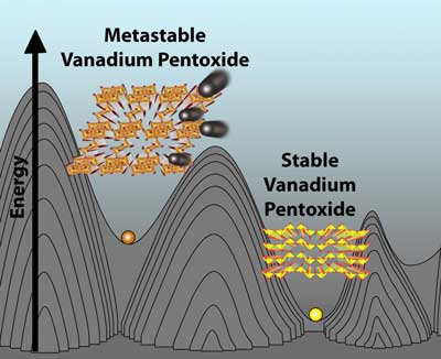
A redesigned metastable phase of vanadium pentoxide (V2O5) shows extraordinary performance as a cathode material for magnesium batteries. The graphic compares the conventional (right) and metastable structures of V2O5. (Image: Justin Andrews, Texas A&M University)
Ironically, the team’s futuristic solution hinges on a redesigned form of an old Li-ion cathode material, vanadium pentoxide, which they proved is capable of reversibly inserting magnesium ions.
„We’ve essentially reconfigured the atoms to provide a different pathway for magnesium ions to travel along, thereby obtaining a viable cathode material in which they can readily be inserted and extracted during discharging and charging of the battery,” Banerjee says.
This rare phenomenon is achieved by limiting the location of the magnesium ions to relatively uncomfortable atomic positions by design, based on the way the vanadium pentoxide is made – a property known as metastability. This metastability helps prevent the magnesium ions from getting trapped within the material and promotes complete harvesting of their charge-storing capacity with negligible degradation of the material after many charge-recharge cycles.

The Ins and Outs of Intercalation

Banerjee, a Davidson Professor of Science in the Texas A&M Department of Chemistry and an affiliated faculty member in the Department of Materials Science and Engineering, has been working for a number of years to better understand ion intercalation – the critical process by which ions like lithium and magnesium move in and out of other materials within intercalation batteries.
Using one of the world’s most powerful soft X-ray microscopes – the Scanning Transmission X-ray Microscope (STXM) and X-ray Emission beamlines – at the Canadian Light Source in tandem with one of the world’s highest resolution aberration-corrected transmission electron microscopes housed at the University of Illinois at Chicago (UIC), Banerjee and collaborators from the Lawrence Berkeley National Laboratory, the UIC and Argonne National Laboratory were able to observe the unique electronic properties of their novel vanadium pentoxide and directly prove magnesium-ion intercalation into the material. Collectively, the team applied decades of combined experience in materials science to explain the fundamental reasons why this new type of vanadium pentoxide is superior to the old version as well as to Li-ion batteries.
Laptops and cell phones are two examples of the many technologies enabled by the rapid development of the lithium-ion battery, which revolutionized energy storage capacity and rechargeability in comparison to its lead-acid and nickel-metal hydride predecessors. However, given the widespread use of lithium not only in portable electronic devices but increasingly in the much larger batteries required for electric vehicles and grid energy storage, lithium is expected to be in increasingly short supply in the long-term. Furthermore, Li-ion batteries are a risky game, as highlighted by recent widely publicized reports detailed in Scientific American, Reuters and Forbes, for example, in which Li-ion-powered devices have either caught fire or exploded as a result of the fundamental flammability and reactivity of lithium.
„Apart from being much safer for consumer applications, magnesium-ion technology is appealing fundamentally because each magnesium ion gives up two electrons per ion – twice the charge, whereas each lithium ion gives up only one,” says Texas A&M chemistry graduate student and NASA Space Technology Research Fellow Justin Andrews, first author on the team’s paper. „This means that, all other considerations aside, if you can store as much magnesium in a material as you can store lithium, you immediately almost double the capacity of the battery.”

Double the Capacity, Double the Trouble

But for all their perceived advantages, magnesium batteries have proven too good to be true since they were first proposed in the 1990s and essentially sidelined by a variety of problems; primarily, the lack of a suitable cathode, or positive electrode – otherwise known as the part of a battery where the magnesium ions enter during discharge of the battery to power an electronic device and then exit during charging.
„Indeed, the most exciting thing about magnesium ions – namely, that they store twice the charge in battery applications – also forms the basis for the biggest challenge,” says collaborating UIC chemist Jordi Cabana. „The higher charge of the magnesium ions make them ‘stick’ much more strongly with surrounding atoms.”
In other words, Banerjee says, the magnesium ions get waylaid as they are traversing through the paths within the cathode material. Their sluggish movement is what makes it so difficult to make viable magnesium batteries.
„In many structures, some of these interactions are very favorable, meaning that the magnesium is quite happy to sit and stay a while in those specific sites,” Andrews explains. „In our material, the magnesium is ‘frustrated’ as it moves through the lattice, because it encounters many less-than-optimal environments. In this sense, it is more than happy to just keep moving right along, leading to an improvement in capacity and diffusion.”
The team’s National Science Foundation-funded research features two additional current and former Texas A&M graduate students, Abhishek Parija and Peter M. Marley, respectively. David Prendergast, a Facility Director at Berkeley Lab’s Molecular Foundry, a U.S. Department of Energy National User Facility for Nanoscale Science Research, helped the Texas A&M team design and interpret their calculations, which were experimentally verified in part by Fakra using Berkeley Lab’s Advanced Light Source along with structural data collected at Argonne National Lab’s Advanced Photon Source. Atomic resolution images of the new form of vanadium pentoxide were collected in collaboration with UIC physicist Robert F. Klie and physics graduate student Arijita Mukherjee and show direct evidence of magnesium intercalated within the material. Battery measurements that show reversibility and confirm the robustness of the cathode material complete the story and were conducted in collaboration with Cabana and former Cabana group member Hyun Deog Yoo.
„On paper, magnesium batteries are highly desirable because they promise greater energy density on top of the ability to solve several of the key issues researchers – and unfortunately consumers – are discovering with lithium-ion batteries, including cost, safety, and performance at the most fundamental levels,” Andrews says. „But the shift from lithium- to magnesium-ion technologies is not straightforward, and the many problems encountered when designing magnesium-ion cathodes have stymied the development of these more sustainable and safer batteries.”

Working Toward a Safer Energy Future

Andrews says the team’s research marks an important turning point in the field because it represents a significant advance toward solving the cathode problem while also highlighting the inherent advantages of using much more imaginative, metastable materials like this new form of vanadium pentoxide. But even he admits there’s much more work to do before this particular ’90s trend comes back in vogue.
„While this research has provided a great deal of insight, there are still several other fundamental problems to overcome before magnesium batteries become a reality,” Andrews adds. „Nevertheless, this work moves magnesium batteries one step closer to reality – namely, a reality where batteries would be less-expensive, lighter and safer for allowing for easier adoption to large-area formats necessary for electric vehicles and to store energy generated by solar and wind sources.”
Source: Texas A&M University

Ceramica de vindecare cu auto-spargere, inspirată de oase, capabilă de recuperare rapidă(Bone-inspired self-crack-healing ceramics capable of quick full strength recovery)

Ceramica auto-vindecată a fost descoperită de un grup de cercetare YNU în 1995. Ușoare și rezistente la căldură, au atras atenția globală pentru utilizarea lor potențială ca materiale de turbină în motoarele de avioane. grupul de cercetare intenționează să dezvolte produse ceramice inovatoare, rezistente la căldură, care nu se vor rupe nici măcar atunci când sunt crăpate prin selectarea unor faze eficiente de vindecare a vindecătorului și, prin urmare, manipularea precisă a capacității de auto-vindecare a ceramici

Posted: Feb 05, 2018

Bone-inspired self-crack-healing ceramics capable of quick full strength recovery

(Nanowerk News) A NIMS-YNU research group discovered that self-healing ceramics undergo three healing stages analogous to bone healing processes: inflammation, repair and remodeling. Using clues provided by bone healing mechanisms, the group added a healing activator to crystalline grain boundaries, enabling the ceramic to fully heal cracks in as little as one minute at 1,000°C, the operating temperature of an aircraft engine (Scientific Reports, „A Novel Design Approach for Self-Crack-Healing Structural Ceramics with 3D Networks of Healing Activator”).

Self-healing process in ceramics
(Left and middle) Self-healing process in ceramics, which completes in as little as one minute. (Right) A network of manganese oxide (green) facilitates self-healing (Image: NIMS)
Self-healing ceramics were discovered by an YNU research group in 1995. Lightweight and heat-resistant, they have been drawing global attention for their potential use as turbine materials in aircraft engines.
However, their self-healing mechanisms had been poorly understood, and cracks only fully healed within a limited temperature range between 1,200 and 1,300°C. Therefore, identification of the mechanism and development of ceramics capable of rapid self-healing within a larger temperature range have long been sought.
The present research group found that self-healing processes occur in three stages: when a ceramic cracks, oxygen enters though the crack and reacts with silicon carbide—a ceramic component—to form silicon dioxide (the inflammation stage). Alumina—a base ceramic material—and silicon dioxide then react to form a gap filling material, sealing the crack (the repair stage).
Finally, the filler crystallizes to restore the original strength in the cracked part (the remodeling stage). In addition, based on the insight that the bodily fluid network in humans promotes the healing of damaged bones, the research group added a trace amount of a healing activator – manganese oxide – to alumina grain boundaries as a 3D network structure.
As a result, newly developed ceramics healed cracks in as little as approximately one minute at 1,000°C, compared to conventional ceramics, which heal cracks in 1,000 hours at 1,000°C.
Based on these results, the research group plans to develop innovative, heat-resistant ceramics which will never break even when cracked by selecting effective healing activator phases and thereby precisely manipulating the self-healing capability of ceramics.
This project was carried out by a research team led by Senior Researcher Toshio Osada (Research Center for Structural Materials (RCSM), NIMS), Group Leader Toru Hara (RCSM, NIMS), Principal Researcher Taichi Abe (RCSM, NIMS), Takahito Ohmura (Deputy Director of the RCSM, NIMS), Chief Researcher Masanori Mitome (International Center for Materials Nanoarchitectonics, NIMS) and Professor Wataru Nakao (Faculty of Engineering, YNU).
Source: National Institute for Materials Science

Respirătoare, electronice portabile pe piele pentru monitorizarea sănătății pe termen lung(Breathable, wearable electronics on skin for long-term health monitoring)

Un senzor electronic hipoalergenic poate fi purtat pe piele continuu timp de o saptamana fara disconfort si este atat de usor si de subtire incat utilizatorii uita ca au chiar si ei, spune un grup de cercetatori de la Universitatea din Tokyo si colaboratorii lor. Electrodul elastic, construit din ochiuri de dimensiuni reduse la nivel de nanoparticule, promite dezvoltarea unor dispozitive neinvazive de e-skin, care pot monitoriza continuu starea de sănătate a unei persoane pe o perioadă lungă de timp ( Nature Nanotechnology , „Inflammation free, permeable gas, lightweight, stretchable on-skin electronics cu nanomeshes ” )

Posted: Feb 05, 2018

Breathable, wearable electronics on skin for long-term health monitoring

(Nanowerk News) A hypoallergenic electronic sensor can be worn on the skin continuously for a week without discomfort, and is so light and thin that users forget they even have it on, says a group of researchers at the University of Tokyo and their collaborators. The elastic electrode constructed of breathable nanoscale meshes holds promise for the development of noninvasive e-skin devices that can monitor a person’s health continuously over a long period (Nature Nanotechnology, „Inflammation-free, gas-permeable, lightweight, stretchable on-skin electronics with nanomeshes”).

Nanomesh conductor applied to back of hand
Nanomesh conductor applied to back of hand. The nanomesh electrode closely follows the contours of the back of the hand for a perfect fit when attached to the skin. The stretchable device lets the skin breathe naturally, and is so thin and light that wearers forget they even have it on. (Image: Someya Laboratory)
Wearable electronics that monitor heart rate and other vital health signals have made headway in recent years, with next-generation gadgets employing lightweight, highly elastic materials attached directly onto the skin for more sensitive, precise measurements.
However, although the ultrathin films and rubber sheets used in these devices adhere and conform well to the skin, their lack of breathability is deemed unsafe for long-term use: dermatological tests show the fine, stretchable materials prevent sweating and block airflow around the skin, causing irritation and inflammation, which ultimately could lead to lasting physiological and psychological effects.
„We learned that devices that can be worn for a week or longer for continuous monitoring were needed for practical use in medical and sports applications,” says Professor Takao Someya at the University of Tokyo’s Graduate School of Engineering whose research group had previously developed an on-skin patch that measured oxygen in blood.
In the current research, the group developed an electrode constructed from nanoscale meshes containing a water-soluble polymer, polyvinyl alcohol (PVA), and a gold layer—materials considered safe and biologically compatible with the body. The device can be applied by spraying a tiny amount of water, which dissolves the PVA nanofibers and allows it to stick easily to the skin—it conformed seamlessly to curvilinear surfaces of human skin, such as sweat pores and the ridges of an index finger’s fingerprint pattern.
The researchers next conducted a skin patch test—for irritations and skin allergies—on 20 subjects and detected no inflammation on the participants’ skin after they had worn the device for a week. The group also evaluated the permeability, with water vapor, of the nanomesh conductor—along with those of other substrates like ultrathin plastic foil and a thin rubber sheet—and found that its porous mesh structure exhibited superior gas permeability compared to that of the other materials.
Furthermore, the scientists proved the device’s mechanical durability through repeated bending and stretching, exceeding 10,000 times, of a conductor attached on the forefinger; they also established its reliability as an electrode for electromyogram recordings when its readings of the electrical activity of muscles were comparable to those obtained through conventional gel electrodes.
„It will become possible to monitor patients’ vital signs without causing any stress or discomfort,” says Someya about the future implications of the team’s research. In addition to nursing care and medical applications, the new device promises to enable continuous, precise monitoring of athletes’ physiological signals and bodily motion without impeding their training or performance.
Source: University of Tokyo