Traductoare piezoelectrice ultrasonice noi concepute pentru aplicații de 360 ​​de grade ( New Spherical Ultrasonic Piezo Transducers Designed for 360 Degree Applications )

PI este un producător de frunte al echipamentelor de control al mișcării de precizie, a motoarelor piezo, a etapelor de transport aerian și a cinematicelor paralele hexapod pentru aplicații semiconductoare, fotoni, bio-nanotehnologie și inginerie medicală. PI a dezvoltat și produce standarde și produse personalizate de precizie cu piezoceramică și unități electromagnetice timp de 4 decenii. Compania a fost certificată ISO 9001 din 1994 și oferă soluții inovatoare, de înaltă calitate pentru OEM și cercetare.
În calitate de lider în traductoarele piezo, mecanismele piezo și soluțiile de control al mișcării de precizie, PI (Physik Instrumente) oferă o serie de emisfere electracerice standard și personalizate și sfere goale pentru aplicații de la ecolocație (sonar) la măsurarea debitului.

Posted: Jun 09, 2016

New Spherical Ultrasonic Piezo Transducers Designed for 360 Degree Applications

(Nanowerk News) As a leader in piezo transducers, piezo mechanisms, and precision motion control solutions, PI (Physik Instrumente) offers a line of standard and custom electro-ceramic hemispheres and hollow spheres for applications from echolocation (sonar) to flow measurement. As a global leader in piezo actuators and sensors, these new components join the existing extensive catalog of disk, rods, cylinders, plates, blocks, rings, and tubes employed by high-tech research and industry fields, including medical engineering, mechanical engineering, aerospace, and semi-conductor applications.
Spherical and hemispherical piezo ultra-sound transducers
Spherical and hemispherical piezo ultra-sound transducers.
Inverse Piezo Effect Applications
Piezo ceramics convert electrical power into mechanical power and vice versa; the inverse piezo effect is harnessed for its fast, precise motion and oscillation generation. For ultrasonic applications that require high bandwidth, hemispheres and hollow spheres are currently available with an outside diameter range from 10 – 60mm and 1mm wall thickness. Uses for electro-ceramic transducers range from the necessary and mundane to out-of-this-world scientific advancement: removing dental plaque, ultrasonic imaging, monitoring flow measurement or fill levels, preparing soil samples on Mars for spectroscopy.
Watch Video of Different Types of Piezo Motor Designs.
Customizable
Made from ferroelectric soft or hard piezo materials, components can be custom-designed to match the requirements of the individual application with respect to the coupling factor and acoustic impedance.
Read Examples of Ultrasonic Piezo Technology.
Standard and Custom
PI has in-house engineered solutions with over 4 decades of experience working with customers to provide products that meet application demands, and can quickly modify existing product designs or provide a fully customized OEM part to fit the exact requirements of the application.
About PI
PI is a leading manufacturer of precision motion control equipment, piezo motors, air bearing stages and hexapod parallel-kinematics for semiconductor applications, photonics, bio-nano-technology and medical engineering. PI has been developing and manufacturing standard & custom precision products with piezoceramic and electromagnetic drives for 4 decades. The company has been ISO 9001 certified since 1994 and provides innovative, high-quality solutions for OEM and research. PI is present worldwide with eleven subsidiaries, R&D / engineering on 3 continents and total staff of 850.
Source: PI (press release)

Read more: New Spherical Ultrasonic Piezo Transducers Designed for 360 Degree Applications

Origami stele Ninja inspiră design nou baterie(Origami Ninja star inspires new battery design )

O baterie de unică folosință care se îndoaie ca o stea ninja de origami ar putea alimenta biosenzori și alte dispozitive mici pentru utilizare în condiții de teren dificile.Baterie de origami pe suport de hârtie.

Posted: Jun 07, 2016

Origami Ninja star inspires new battery design

(Nanowerk News) A new disposable battery that folds like an origami ninja star could power biosensors and other small devices for use in challenging field conditions, says an engineer at Binghamton University, State University of New York.

Seokheun “Sean” Choi, assistant professor of computer and electrical engineering at Binghamton University, along with two of his students, developed the device, a microbial fuel cell that runs on the bacteria available in a few drops of dirty water. They report on their invention in a new paper published online in the journal Biosensors and Bioelectronics („A disposable power source in resource-limited environments: A paper-based biobattery generating electricity from wastewater”).
disposable battery that folds like an origami ninja star
A new disposable battery that folds like an origami ninja star could power biosensors and other small devices for use in challenging field conditions.
Choi previously developed a paper-based origami battery. The first design, shaped like a matchbook, stacked four modules together. The ninja star version, which measures about 2.5 inches wide, boasts increased power and voltage, with eight small batteries connected in series.
“Last time, it was a proof of concept. The power density was in the nanowatt range,” said Choi. “This time, we increased it to the microwatt range. We can light an LED for about 20 minutes or power other types of biosensors.”
Paper-based biosensors include pregnancy tests and HIV tests. The sensitivity of such tests is limited, Choi said, and a battery like his could allow the use of more sophisticated fluorescent or electrochemical biosensors in developing countries. “Commercially available batteries are too wasteful and expensive for the field,” he said. “Ultimately, I’d like to develop instant, disposable, accessible bio-batteries for use in resource-limited regions.”
The new design folds into a star with one inlet at its center and the electrical contacts at the points of each side. After a few drops of dirty water are placed into the inlet, the device can be opened into a Frisbee-like ring to allow each of the eight fuel cells to work. Each module is a sandwich of five functional layers with its own anode, proton exchange membrane and air-cathode.
Choi’s original matchbook-sized battery could be produced for about 5 cents. The new ninja star device is more expensive — roughly 70 cents — in part because it uses not only filter paper but also carbon cloth for the anode as well as copper tape. The team’s next goal is to produce a fully paper-based device that has the power density of the new design and a lower price tag.
Choi prides himself on involving students in his research, and this paper represents a special triumph for co-author Landen Kwan, who worked on the project during a National Science Foundation Research Experiences for Undergraduates program at Binghamton in the summer of 2015. Kwan, then a student at Queensborough Community College, is now enrolled at Stony Brook University. Binghamton doctoral student Arwa Fraiwan also contributed to the study, which was funded by the National Science Foundation.
Source: Binghamton University

Read more: Origami Ninja star inspires new battery design

Undă moleculară a catalizatorului pe bază de oxid de metal nanosizat

Studiul recent al grupului Ananikov a vizat formarea legăturii carbon-heteroatom catalizată de oxizii de cupru de dimensiuni nano. Acest tip de reacție catalitică interesantă utilizează nanoparticule CuO și Cu2O pentru a promova transformările chimice. Studiul mecanic a relevat o imagine destul de complicată a evoluției catalizatorului. În reacția C-S de cuplare încrucișată, oxidul de cupru cu dimensiunea nano a fost transformat în specii metalice solubile: tiolații de cupru [Cu (SR) n] s-au format ușor în reacția oxidului de cupru cu tioli (RSH). Într-adevăr, procesul studiat demonstrează că reacția presupusă în mod tradițional pe suprafața oxidului de metal nu aduce o contribuție primară la formarea produsului. Modificarea oxidului metalic și transferul de soluții corespunzătoare de tiolat de metal în soluție au fost responsabile de formarea formei active a catalizatorului.
În ciuda căii catalitice eterogene așteptate, studiul reacției de cuplare încrucișată a evidențiat solubilizarea oxidului de cupru și a catalizei omogene de stare a soluției. Mișcarea complecșilor de cupru la soluție a fost însoțită de eliberarea apei: Cu2O + 2RSH = 2CuSR + H2O

Posted: Jun 01, 2016

Molecular wave of nanosized metal oxide catalyst

(Nanowerk News) Metal oxides represent primary source of catalytic materials for a variety of industrial and research applications, either as an active phase or as a supporting phase. Mechanistic studies conducted by Ananikov group revealed formation of catalytically active species due to partial dissolution of metal oxide in the reaction mixture and generation of soluble metal complexes (ACS Catalysis, „Nature of the Copper-Oxide-Mediated C–S Cross-Coupling Reaction: Leaching of Catalytically Active Species from the Metal Oxide Surface”). Therefore, metal oxides are not directly involved in heterogeneous catalysis; rather, they can be considered as a source of reactive species for solution-state homogeneous catalysis (DOI: 10.1021/acscatal.6b00337).

Catalysis is the major driving force in modern industry. Catalysis is vital in production of fuels, materials, drugs, and a variety of molecular and composite chemicals. Around 80% of all industrial processes involve catalytically produced components or catalytic technologies. Metal oxides represent a traditional class of solid materials to create active phases of catalysts or to construct supports for other types of active centers. Metal oxides as active phases are designed to render chemical transformation and directly participate in the catalytic cycle. Metal oxides as supporting phases are used to introduce innocent component with high stability.
Cryogenic scanning electron microscopy study of solution phase of catalytic cross-coupling reaction
Cryogenic scanning electron microscopy study of solution phase of catalytic cross-coupling reaction. (Image: Ananikov group)
Recent study of Ananikov group addressed carbon-heteroatom bond formation catalyzed by nano-sized copper oxides. This intriguing type of catalytic reactions utilizes CuO and Cu2O nanoparticles to promote chemical transformations. Mechanistic study revealed rather complicated picture of catalyst evolution. In C-S cross-coupling reaction, nano-sized copper oxide was transformed to soluble metal species: copper thiolates [Cu(SR)n] were readily formed in the reaction of copper oxide with thiols (RSH). Indeed, the studied process demonstrates that traditionally assumed reaction on the surface of metal oxide do not make a primary contribution to the product formation. Modification of metal oxide and transfer of corresponding metal thiolate species to solution were responsible for the formation of active form of the catalyst.
Despite expected heterogeneous catalytic pathway, the study of cross-coupling reaction revealed solubilization of copper oxide and solution state homogeneous catalysis. Movement of copper complexes to solution was accompanied by release of water: Cu2O + 2RSH = 2CuSR + H2O. Cryogenic scanning electron microscopy suggested formation of a complex catalytic system with substantial evolution of initial form of metal oxide during the catalytic reaction (see Figure).
The occurred transformations may change the paradigm of metal-oxide-based systems application in catalysis for both options – active phase and supporting phase. In the former option, if metal oxide is assumed as an active phase, transition to solution drastically changes the nature of the catalytic process. In the latter option, if metal oxide is utilized as a support, the oxide does not behave as an innocent material and cannot be recovered in the original state.
Catalyst stability and recycling are the key properties affected by the reported transformation of the metal oxide. Knowledge of mechanistic nature of catalyst evolution opens up new possibilities to reduce cost of chemicals production processes.
Source: Zelinsky Institute of Organic Chemistry
Subscribe to a free copy of one of our daily
Nanowerk Newsletter Email Digests
with a compilation of all of the day’s news.

Read more: Molecular wave of nanosized metal oxide catalyst

Nanotehnologii sunt pași mici spre fabricarea atomică la scară 3D

se va deschide porțile pentru tehnologiile emergente în calcul, energie, cataliza, și o serie de alt articol de revizuire recente fields.A (în ACS Nano „Directing Materia: Spre Nanofabrication Atomic-Scale 3D „) Ilustrează utilizarea fascicule de electroni și ioni concentrat (e-grinzi și traverse) pentru a induce reacții chimice foarte localizate la interfețe solide la vapori și solid-lichid, amorfe transformări de fază cristaline cu precizie strat atomic, iar mișcarea atom specific dopant unic, în barilor de cristal, punând astfel bazele pentru asamblarea atomically precis direcționată a materialelor și a creșterii devices.

Nanotechnology’s tiny steps toward atomic-scale 3D fabrication

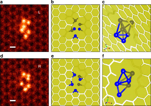
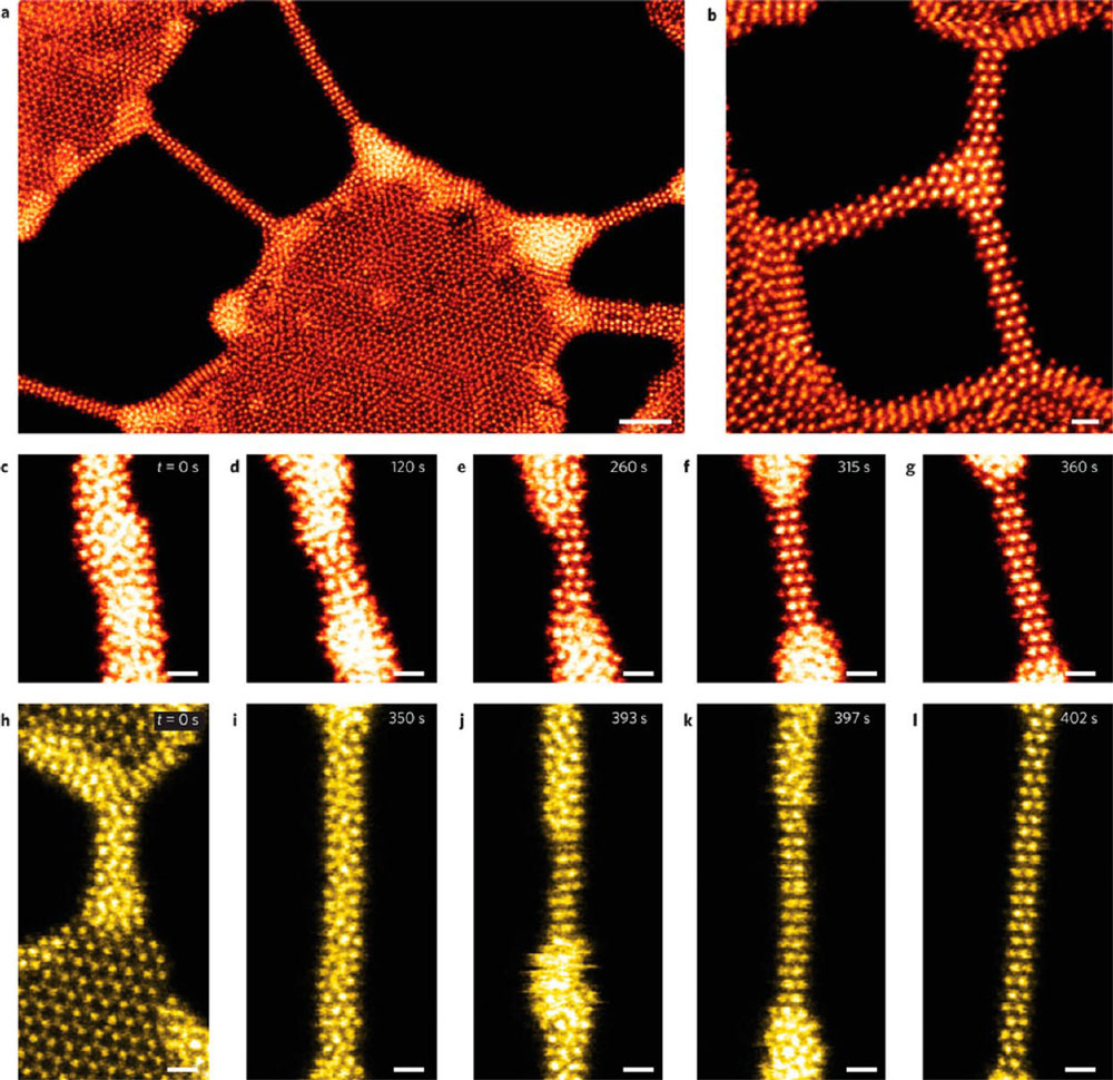
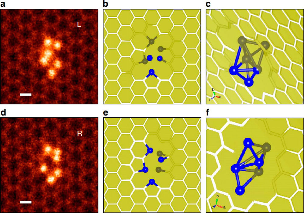
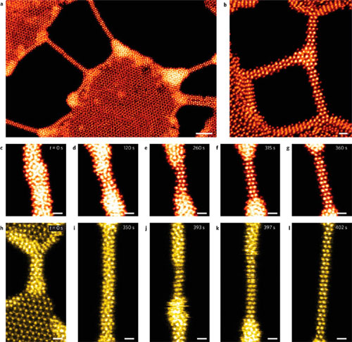
(Nanowerk Spotlight) Over the past few decades, the development of electron microscopy has gone hand in hand with techniques for atomically precise fabrication of 3D structures based on electron and ion beams.All material functionality is governed by the type, arrangement, and bonding of atoms. Enabling the capacity to perform atomically precise directed assembly of matter will open the doors to emerging technologies in computing, energy, catalysis, and a plethora of other fields.A recent review article in ACS Nano („Directing Matter: Toward Atomic-Scale 3D Nanofabrication”) illustrates the use of focused electron and ion beams (e-beams and i-beams) to induce highly localized chemical reactions at solid-vapor and solid-liquid interfaces, amorphous to crystalline phase transformations with atomic layer precision, and the motion of specific single dopant atoms within crystal lattices, thus laying the foundation for atomically precise directed assembly of materials and devices.The growth of plasmonic gold nanostructures by electron beam induced deposition (EBID) has been demonstrated already 10 years ago and the physics behind this nanofabrication technique has been explored (see for instance: „Scientists unveil new physics behind the nanofabrication technique of electron beam induced deposition„). The process has been improved over the years and researchers are now able to grow structures much faster with a broad range of precursors (see: „A new process for rapidly fabricating complex 3D nanostructures„).In their review, the authors emphasize the synergy between imaging, nanofabrication, and modeling in order to reveal the fundamental mechanisms of charged particle-surface interaction.They present several recent examples that suggest the feasibility of atomically precise fabrication in the bulk, including creating specific dopant configurations, ordered vacancy planes, and other structures.The article covers EBID/IBID from gas or liquid precursors, e-beam-induced crystallization in the bulk, and the ability to image and to analyze single atoms and defects in 2D structures.Patterning of a MoSe nanowire network(a) Patterning of a MoSe nanowire network, where each nanowire is sculpted individually. (b) Ramified Y junction made of three MoSe nanowires. (c-l) Serial snapshots of the sculpting process of individual MoSe (c-g) and MoS (h-l) nanowires. All images are STEM Z-contrast images, false-colored for better visibility. Scale bars, 2 nm (a), 0.5 nm (b-l). (© Nature Publishing Group) (click on image to enlarge)Electron-beam/ion-beam-induced deposition from gas precursorsDiscussing Focused Electron-Beam-Induced Processing – a nanoscale direct-write synthesis technique where an electron beam directs assembly of a surface-adsorbed precursor via an electron-stimulated chemical reaction – and Focused Ion-Beam-Induced Processing – where the deposition and etching of nanoscale materials is induced by a focused ion beam – the authors show how these techniques were developed; they discuss their benefits but also obstacles to large-scale implementation.Eelectron-beam/ion-beam-induced deposition from liquid precursorsAn alternative pathway to using gas precursors for direct nanofabrication is direct-write with electron and/or ion beams with a liquid precursor. This method utilizes the e-beam or i-beam as an ionizing source that decomposes solvent molecules to generate solvated electrons and other radicals in a complex cascade of reaction pathways.When using electrons to direct-write in solution, the minimal size of the created structures is limited to the nanometer scale due to the fundamental physics of the interactions between the electron beam and the liquid, including the lateral transport of solvated electrons and ionic species. The helium ion beam, on the other hand, might offer the potential for improved resolution.Atomically precise e-beam-induced crystallization in the bulkE-beam and i-beam patterning are popular and versatile approaches for creating structures on ∼10-100 nm scales, typically in SEM-like geometry. However, in the field of imaging, high-resolution transmission electron microscopy (TEM) has long been a tool of choice because it allows for atomic-scale studies of crystals and nanoparticles.As the authors note, over the last 15 years, aberration correction has pushed the achievable resolution into the sub-angstrom range and allowed for much greater localization of the probe in STEM geometry, suggesting that higher precision patterning should be achievable using these improved electron probes.Provided that the electron beam path can be programmed, localization of features in the areas illuminated by the beam and the ability to control their growth allows for more complex patterning.The team writes that the manufacturing of epitaxial crystalline nanostructures with predefined shapes is particularly interesting, as adjustable strain states could yield ways to control properties, and the ordered final structures can be more easily modeled than amorphous structures.Si6 cluster configurations embedded in a graphene pore(a,d) ADF Z-contrast images of two representative stable Si6 cluster configurations embedded in a graphene pore. (b,e) Two- and (c,f) three-dimensional structures obtained from first-principles calculations. Si and C atoms below the graphene plane are shaded. L or R labels are used to highlight when the oscillating Si atom is either at the left or right sites. Scale bar, 0.2 nm. (© Nature Publishing Group) (click on image to enlarge)Single-atom dynamics and beyondThe difficulties in manipulating single atoms for nanoscale engineering were famously highlighted by Stemmer („Three-dimensional imaging of individual dopant atoms in SrTiO3„).The use of an electron beam might provide an answer to this dilemma by allowing the direct addressing of single atoms in bulk 3D materials.The review highlights results („Direct Observation of Dopant Atom Diffusion in a Bulk Semiconductor Crystal Enhanced by a Large Size Mismatch”) that show the interplay between the materials science aspect of calculated diffusion barriers and the energy supplied to the sample by the electron beam.As the authors point out: „This relation is important because it is possible to directly control the accelerating voltage in a (S)TEM, thereby altering the energy transferred to the material. This concept raises several tantalizing prospects for the future as to what extent the final position of the dopant can be controlled. If single dopants could be directly manipulated, an obvious application would be toward quantum computing.”Link theory and dataIncreasing computer power and more sophisticated analytical software allows analyzing experimental data in near real-time. This provides the ability to have rapid feedback to and from models and simulations that both inform and validate the simulations/model, which may ultimately provide predictive capabilities to guide experiments.Until recently, advances in microscopy relied on improvements in the instrumentation platforms, focused probes, lenses, detectors, measurement modalities, and mathematical tools for extraction of materials-specific parameters from imaging data. However, in almost all cases, due to the complexity and size of the available signals, only a small, manually selected subset of the fully generated information volume is recorded and an even smaller subset of this is processed.In the future, new tools will allow the (real-time) monitoring and analysis of the full data stream from the microscope. This would also allow researchers to take full advantage of all of the information contained in experimental data to provide input into computational methods to predict and understand materials.The authors conclude that „in order to see what is happening as it is happening, a significant emphasis must be placed on forming a tight loop between expansive dynamic streaming EELS (electron energy loss spectroscopy), tomographic, and ptychographic data sets provided by experimental platforms and the model-driven interpretation of the data. As the techniques mature, these theory-in-the-loop approaches will also significantly expedite the quality control process, an important facet in transitioning from scientific test-beds to high-volume manufacturing.”By Michael is author of two books by the Royal Society of Chemistry: Nano-Society: Pushing the Boundaries of Technology and Nanotechnology: The Future is Tiny.
Read more: Nanotechnology’s tiny steps toward atomic-scale 3D fabrication

Sprijinirea dezvoltării responsabile a nanotehnologiei

Se pot detecta acum nanoparticule cu mult sub nivelurile de toxicitate cunoscute și sub pragul acțiunilor de reglementare economice și rezonabile Înlătură probleme complexe de determinare a biomarkerilor expunerii legate de boală problemei complexe de determinare a biomarkerilor expunerii legate de boală.

Posted: May 31, 2016

Supporting responsible development of nanotechnology

(Nanowerk News) Nanotechnology environmental, health, and safety (nanoEHS) activities have become a hallmark of the National Nanotechnology Initiative (NNI). Well-coordinated nanoEHS research and development (R&D) is essential to ensuring a future in which responsible development of nanotechnology provides maximum benefit to society and the environment. These efforts are pivotal in contributing to American innovation and to advancing manufacturing and economic competitiveness.

Progress in nanoEHS research is vital to establishing the regulatory certainty and public confidence needed for companies to bring their nanotechnology products to market and to ensuring that these products are safe and sustainable throughout their life cycles.
Three reports recently released highlight Federal investments and activities in this area, progress and needs in understanding exposure from consumer products, and how businesses can protect their nanotechnology workforce. In addition, an upcoming workshop will explore the work of joint U.S.–European Union nanoEHS communities of research in developing a shared repertoire of protocols and methods.
The recently released NNI Supplement to the President’s Budget for Fiscal Year 2017, which serves as the annual report for the NNI, highlights the programs and coordinated activities taking place across the many departments, independent agencies, and commissions participating today in the NNI—an initiative that continues to serve as a model for effective coordination of Federal science and technology R&D.
As detailed in this report, nanoEHS activities continue to account for about 10 percent of the annual NNI budget, with cumulative Federal R&D investments in this area exceeding $1 billion over the past decade. This report includes descriptions of a wide variety of individual agency and coordinated activities supporting the responsible development of nanotechnology.
To understand and control the risks of using any new materials in consumer products, it is important to understand the potential for exposure and any associated hazards across product life cycles. Last month, the NNI released a report, Quantifying Exposure to Engineered Nanomaterials (QEEN) from Manufactured Products: Addressing Environmental, Health, and Safety Implications, summarizing a workshop on this topic sponsored by the U.S. Consumer Product Safety Commission (CPSC).
The main goals of the workshop were to assess progress in developing tools and methods for quantifying exposure to engineered nanomaterials across the product life cycle, and to identify new research needed to advance exposure assessment for nanotechnology-enabled products. As noted in the report, significant progress has been made in the ability to quantify nanomaterial exposures, such that available methods can now detect nanoparticles well below known toxicity levels and beneath the threshold of economical and reasonable regulatory action. However, greater understanding of exposure risks in “real-world” scenarios is needed, along with techniques, based on alternative testing models and high-throughput methods for rapidly estimating exposures.
The technical experts who participated in CPSC’s workshop recommended that future work focus on the complex issue of determining biomarkers of exposure linked to disease, which will require substantive public–private collaboration, partnership, and knowledge sharing. Recognizing these needs, the President’s 2017 Budget request for CPSC (pdf) includes funds for a new nanotechnology center led by the National Institute of Environmental Health Sciences (NIEHS) to develop test methods and to quantify and characterize the presence, release, and mechanisms of consumer exposure to nanomaterials in consumer products. This cost-effective, interagency collaboration will enable CPSC—through NIEHS—to collect the needed data to inform the safety of nanotechnology in consumer products and allow CPSC to benefit from NIEHS’s scientific network and experience.
Managing EHS risks across a product’s lifecycle includes protecting the workers who manufacture those products. The National Institute for Occupational Safety and Health has issued a series of documents providing guidance to this emerging industry, including the recently released publication Building a Safety Program to Protect the Nanotechnology Workforce: A Guide for Small to Medium-Sized Enterprises. This guide provides business owners with the tools necessary to develop and implement a written health and safety program to protect their employees.
The responsible development of nanotechnology is a goal that the United States shares with many countries. The United States and the European Union are engaged in notable cooperation on this front. European and American scientists engaged in nanoEHS research convene annually for a joint workshop to identify areas of shared interest and mechanisms for collaboration to advance nanoEHS science. The 2016 joint workshop will be held on June 6–7, 2016 in Arlington, VA, and is free and open to the public. Registration opened on April 6, 2016, on a first-come, first-served basis.
Source: By Lloyd Whitman, Assistant Director for Nanotechnology and Advanced Materials at the White House Office of Science and Technology Policy

Read more: Supporting responsible development of nanotechnology

Creșterea cristalelor perfecte prin umplerea golurilor

Cristalele sunt materiale solide compuse din blocuri microscopice, dispuse în modele foarte ordonat. Ei au nenumărate aplicații, variind de la metalurgie la electronice la bijuterii. Multe dintre proprietățile au făcut cristale utile depind de modelul detaliat al aranjament al constituenților lor, care, la rândul său, este extrem de sensibil la detaliile interacțiunii dintre blocurile de construcție. In cristalele moleculare si atomice forțele interparticule sunt fixate de Natura, iar singura modalitate de tuning aranjament microscopic este de a varia fie condițiile externe (temperatură, presiune, etc.) sau de a modifica particulele Înșiși.amestecurilor de coloizi si particule pe baza de polimeri numiti polimeri stele

Posted: May 31, 2016

Growing perfect crystals by filling the gaps

(Nanowerk News) Be it in phyics, metallurgy, gemology or engineering, the applications of crystals are very broad. A research team including Christos Likos and Lorenzo Rovigatti from the Faculty of Physics of the University of Vienna, in collaboration with the National Institute of Standards and Technology (NIST, USA) and Princeton University (USA) has developed a new method to assemble large, periodic crystals. The results have been published in the Journal ACS Nano („Bottom-Up Colloidal Crystal Assembly with a Twist”).

long-range order can be restored by employing mixtures of colloids and polymer-based particles called star polymers
The scientists have shown that long-range order can be restored by employing mixtures of colloids and polymer-based particles called star polymers (Image: Christos Likos, Universität Wien)
Crystals are solid materials composed of microscopic building blocks arranged in highly ordered patterns. They have countless applications, ranging from metallurgy to jewellery to electronics. Many of the properties that make crystals useful depend on the detailed pattern of arrangement of their constituents, which, in turn, is highly sensitive to the details of the interaction between the building blocks. In molecular and atomic crystals the interparticle forces are fixed by Nature, and the only way of tuning the microscopic arrangement is to either vary the external conditions (temperature, pressure, etc.) or change the particles themselves.
By contrast, insoft matter Physics , where the building blocks are orders of magnitude larger and much more complex than atoms, it is possible to design and engineer building blocks with extremely tunable properties. Consequently, much effort has been devoted to the synthesis of colloids that self-assemble into highly symmetric patterns with technologically relevant properties. For instance, there exist specific crystal lattices that exhibit very exciting optical properties, the so-called photonic crystals – periodic structures that allow certain bands of wavelengths of light to propagate through their interior while blocking other ones.
A natural example of a photonic crystal is the opal, whose fascinating coloration is due to the way the light interacts with its microscopic structure of colloidal particles arranged on a regular lattice. The multicolored iridescence of the precious opal, the source of its charming appearance, is due to the presence of several small crystals, known as crystallites, which are randomly oriented with respect to the one another.
In addition, the assembly of colloidal crystals is often confounded by polymorphism: „Different structures are characterised by comparable thermodynamic stabilities, making it difficult to produce a single morphology at will”, says Christos Likos from the Faculty of Physics of the University of Vienna.
The resulting lack of long-range order is detrimental for many applications. Accordingly, strategies need to be developed that enhance the growth of long-ranged, monocrystalline samples in (real or numerical) experiments. Accordingly, scientists have been working hard to deveop strategies that enhance the growth of large, monocrystalline structures. Employing computer simulations, a new method has now been developed that allows the assembly of technologically relevant, non-polymorphic crystals.
„The system crystallises into a mixture of difference micocrystals. However, the competing structures assembled by the colloids have different geometries and different internal void distributions. This difference can be exploited by tuning the size of polymer additive to interact uniquely with the void symmetry of the desired crystal, effectively stabilising it against the competitor”, explains Lise-Meitner Fellow Lorenzo Rovigatti, working at the group of Christos Likos.
The results of the research team serve not only to illustrate an alternative to existing approaches which, in many cases, produce unsatisfactory results, but also to guide experimental realizations of highly-ordered colloidal open crystals in the near future.
Source: Universität Wien

Read more: Growing perfect crystals by filling the gaps

Camera de atracție pentru nanostructuri

O tehnologie care ar putea transfera și de a salva datele codificate pe undele de lumina ar fi de 100.000 de ori mai rapid decât sistemele actuale. O interacțiune materie de lumină, care ar putea deschide calea de a căuta produse electronice bazate pe lumina a fost investigata de catre oamenii de stiinta de la Laboratorul de Attosecond Fizica (PLA) Ludwig-Maximilians-University (LMU) și Institutul Max Planck de Optică Cuantică (MPQ ), în colaborare cu colegii de la Catedra de fizica cu laser de la Universitatea din Erlangen-Nuremberg. Cercetatorii au trimis impulsuri laser intens pe un nanofire mic din aur. Cele ultrascurte impulsului laser excitat vibrația electronilor se deplasează liber în metal.
Acest lucru a dus la electromagnetic „lângă câmpuri“ la suprafața firului. Cvasi-câmpuri a oscilat cu o schimbare de câteva sute de attoseconds în ceea ce privește câmpul cu laser interesant (un Attosecond este o miliardime de miliardime de secundă). Această schimbare ce Măsurat folosind impulsuri de lumina Attosecond pe care oamenii de știință trimis ulterior Onto nanowire.
Atunci când lumina se aprinde metale, aceasta poate duce la lucruri curioase în microcosmos la suprafață. Câmpul electromagnetic al luminii excită vibratii ale electronilor din metal. Această interacțiune determină formarea „aproape-câmpuri“ – câmpuri electromagnetice localizate în apropierea suprafeței metalului.

Posted: May 31, 2016

Attosecond camera for nanostructures

(Nanowerk News) Physicists of the Laboratory for Attosecond Physics at the Max Planck Institute of Quantum Optics and the Ludwig-Maximilians-Universität Munich in collaboration with scientists from the Friedrich-Alexander-Universität Erlangen-Nürnberg have observed a light-matter phenomenon in nano-optics, which lasts only attoseconds („Attosecond nanoscale near-field sampling”).

The interaction between light and matter is of key importance in nature, the most prominent example being photosynthesis. Light-matter interactions have also been used extensively in technology, and will continue to be important in electronics of the future.
When laser light interacts with a nanoneedle (yellow), electromagnetic near-fields are formed at its surface
When laser light interacts with a nanoneedle (yellow), electromagnetic near-fields are formed at its surface. A second laser pulse (purple) emits an electron (green) from the needle, permitting to characterize the near-fields. (Image: Christian Hackenberger)
A technology that could transfer and save data encoded on light waves would be 100.000-times faster than current systems. A light-matter interaction which could pave the way to such light-driven electronics has been investigated by scientists from the Laboratory for Attosecond Physics (LAP) at the Ludwig-Maximilians-Universität (LMU) and the Max Planck Institute of Quantum Optics (MPQ), in collaboration with colleagues from the Chair for Laser Physics at the Friedrich-Alexander-Universität Erlangen-Nürnberg. The researchers sent intense laser pulses onto a tiny nanowire made of gold. The ultrashort laser pulses excited vibrations of the freely moving electrons in the metal.
This resulted in electromagnetic ‘near-fields’ at the surface of the wire. The near-fields oscillated with a shift of a few hundred attoseconds with respect to the exciting laser field (one attosecond is a billionth of a billionth of a second). This shift was measured using attosecond light pulses which the scientists subsequently sent onto the nanowire.
When light illuminates metals, it can result in curious things in the microcosm at the surface. The electromagnetic field of the light excites vibrations of the electrons in the metal. This interaction causes the formation of ‘near-fields’ – electromagnetic fields localized close to the surface of the metal.
How near-fields behave under the influence of light has now been investigated by an international team of physicists at the Laboratory for Attosecond Physics of the Ludwig-Maximilians-Universität and the Max Planck Institute of Quantum Optics in close collaboration with scientists of the Chair for Laser Physics at the Friedrich-Alexander-Universität Erlangen-Nürnberg.
The researchers sent strong infrared laser pulses onto a gold nanowire. These laser pulses are so short that they are composed of only a few oscillations of the light field. When the light illuminated the nanowire it excited collective vibrations of the conducting electrons surrounding the gold atoms. Through these electron motions, near-fields were created at the surface of the wire.
The physicists wanted to study the timing of the near-fields with respect to the light fields. To do this they sent a second light pulse with an extremely short duration of just a couple of hundred attoseconds onto the nanostructure shortly after the first light pulse. The second flash released individual electrons from the nanowire. When these electrons reached the surface, they were accelerated by the near-fields and detected. Analysis of the electrons showed that the near-fields were oscillating with a time shift of about 250 attoseconds with respect to the incident light, and that they were leading in their vibrations. In other words: the near-field vibrations reached their maximum amplitude 250 attoseconds earlier than the vibrations of the light field.
“Fields and surface waves at nanostructures are of central importance for the development of lightwave-electronics. With the demonstrated technique they can now be sharply resolved.”, explained Prof. Matthias Kling, the leader of the team carrying out the experiments in Munich.
The experiments pave the way towards more complex studies of light-matter interaction in metals that are of interest in nano-optics and the light-driven electronics of the future. Such electronics would work at the frequencies of light. Light oscillates a million billion times per second, i.e. with petahertz frequencies – about 100.000 times faster than electronics available at the moment. The ultimate limit of data processing could be reached.
Source: Max Planck Institute of Quantum Optics

Read more: Attosecond camera for nanostructures

Văzând nanofibrele „vii” în timp real

care poseda flexibilitatea si diversitatea de functii care exista intr-o celula vii.Putem controla auto-sortarea cu moleculele artificiale,putem lucra pentru a dezvolta inteligente

Posted: May 31, 2016

Seeing ‘living’ nanofibers in real time (w/video)

(Nanowerk News) The Greek goddess Psyche borrowed help from ants to sort a room full of different grains. Cells, on the other hand, do something similar without Olympian assistance, as they organize molecules into robust, functional fibers. Now scientists are able to see self-sorting phenomena happen in real time with artificial molecules.

The achievement, reported in Nature Chemistry („In situ real-time imaging of self-sorted supramolecular nanofibers”), elucidates how two different types of nanofibers sort themselves into organized structures under artificial conditions.
Real-time imaging of supramolecular nanofibers?Video: Itaru Hamachi)
„Basic cellular structures, such as actin filaments, come into being through the autonomous self-sorting of individual molecules, even though a tremendous variety of proteins and small molecules are present inside the cell,” says lead author Hajime Shigemitsu, a researcher in Itaru Hamachi’s lab at Kyoto University.
„Imagine a box filled with an assortment of building blocks – it’s as if the same type of blocks started sorting themselves into neat bundles all on their own. In living cells, such phenomena always happen, enabling accurate self-assembling of proteins, which is essential for cell functions.”
„If we are able to control self-sorting with artificial molecules, we can work toward developing intelligent, next-generation biomimics that possess the flexibility and diversity of functions that exist in a living cell.”
Study co-author Ryou Kubota explains that previous studies have already made artificial molecules build themselves into fibers – but only when there was one type of molecule around. Having a jumble of types, on the other hand, made the molecules confused.
„The difficulty in inducing self-assembly with artificial molecules is that they don’t recognize the same type of molecule, unlike molecules in the natural world. Different types of artificial molecules interact with each other and make an unsorted cluster.”
From a database of structural analyses, Hamachi and colleagues discovered a combination of nanofibers – namely a peptide-based and lipid-based hydrogelator – that would make sorted fibers without mixing with the other. They then tethered the fibers with fluorescent probes; with a type of microscope typically used in cell imaging, the team was able to observe directly and in real-time how the artificial molecules sorted themselves.
„Ultimately, this finding could help develop new materials that respond dynamically to different environments and stimuli,” elaborates Hamachi. „This insight is not only useful for materials science, but may also provide useful clues for understanding self-organization in cells.”
Source: Kyoto University

Read more: Seeing ‘living’ nanofibers in real time (w/video)

Sondă nanometrica ar putea produce îmbunătățiri majore în baterii și celule de combustie

îmbunătățiri majore în baterii și celule de combustie îmbunătățiri majore în baterii și celule de combustie.Proprietățile materialelor pot afecta performanța bateriilor și a altor sisteme electrochimice.

Posted: May 31, 2016

Tiny probe could produce big improvements in batteries and fuel cells

(Nanowerk News) A team of American and Chinese researchers has developed a new tool that could aid in the quest for better batteries and fuel cells.

Although battery technology has come a long way since Alessandro Volta first stacked metal discs in a „voltaic pile” to generate electricity, major improvements are still needed to meet the energy challenges of the future, such as powering electric cars and storing renewable energy cheaply and efficiently.
The key to the needed improvements likely lies in the nanoscale, said Jiangyu Li, a professor of Mechanical Engineering at the University of Washington in Seattle. The nanoscale is a realm so tiny that the movement of a few atoms or molecules can shift the landscape. Li and his colleagues have built a new window into this world to help scientists better understand how batteries really work. They describe their nanoscale probe in the Journal of Applied Physics („Scanning thermo-ionic microscopy for probing local electrochemistry at the nanoscale”).
nanoscale map of the metal ceria
This is a nanoscale map of the metal ceria produced with the new probe shows a higher response, represented by a yellow color, near the boundary between grains of metal. The higher response corresponds to a higher concentration of charged species. (Image: Ehsan Nasr Esfahani/University of Washington) (click on image to enlarge)
Batteries, and their close kin fuel cells, produce electricity through chemical reactions. The rates at which these reactions occur determine how fast the battery can charge, how much power it can provide, and how quickly it degrades.
Although the material in a battery electrode may look uniform to the human eye, to the atoms themselves, the environment is surprisingly diverse.
Near the surface and at the interfaces between materials, huge shifts in properties can occur – and the shifts can affect the reaction rates in complex and difficult-to-understand ways.
Research in the last ten to fifteen years has revealed just how much local variations in material properties can affect the performance of batteries and other electrochemical systems, Li said.
The complex nanoscale landscape makes it tricky to fully understand what’s going on, but „it may also create new opportunities to engineer materials properties so as to achieve quantum leaps in performance,” he said.
To get a better understanding of how chemical reactions progress at the level of atoms and molecules, Li and his colleagues developed a nanoscale probe. The method is similar to atomic force microscopies: A tiny cantilever „feels” the material and builds a map of its properties with a resolution of nanometers or smaller.
In the case of the new electrochemical probe, the cantilever is heated with an electrical current, causing fluctuations in temperature and localized stress in the material beneath the probe. As a result, atoms and ions within the material move around, causing it to expand and contract. This expansion and contraction causes the cantilever to vibrate, which can be measured accurately using a laser beam shining on the top of the cantilever.
If a large concentration of ions or other charged particles exist in the vicinity of the probe tip, changes in their concentration will cause the material to deform further, similar to the way wood swells when it gets wet. The deformation is called Vegard strain.
Both Vegard strain and standard thermal expansion affect the vibration of the material, but in different ways. If the vibrations were like musical notes, the thermally-induced Vegard strain is like a harmonic overtone, ringing one octave higher than the note being played, Li explained.
The device identifies the Vegard strain-induced vibrations and can extrapolate the concentration of ions and electronic defects near the probe tip. The approach has advantages over other types of atomic microscopy that use voltage perturbations to generate a response, since voltage can produce many different kinds of responses, and it is difficult to isolate the part of the response related to shifts in ionic and electronic defect concentration. Thermal responses are easier to identify, although one disadvantage of the new system is that it can only probe rates slower than the heat transfer processes in the vicinity of the tip.
Still, the team believes the new method will offer researchers a valuable tool for studying electrochemical material properties at the nanoscale. They tested it by measuring the concentration of charged species in Sm-doped ceria and LiFePO4, important materials in solid oxide fuel cells and lithium batteries, respectively.
„The concentration of ionic and electronic species are often tied to important rate properties of electrochemical materials – such as surface reactions, interfacial charge transfer, and bulk and surface diffusion – that govern the device performance,” Li said. „By measuring these properties locally on the nanoscale, we can build a much better understanding of how electrochemical systems really work, and thus how to develop new materials with much higher performance.”
Source: American Institute of Physics

Read more: Tiny probe could produce big improvements in batteries and fuel cells

Corpurile rapide, elastice ar putea da naștere unui nou val de electronice portabile

Înlătură termen: dimensiuni ale lungimii de undă cuprinse între un milimetru și un metru dimensiuni ale lungimii de undă cuprinse între un milimetru și un metruÎnlătură termen: frecvențele radio cu microunde sunt unde electromagnetice care utilizează frecvențe în intervalul de la 3 gigahertzi la 300 gigahertzi. Aceasta se încadrează direct în domeniul 5G frecvențele radio cu microunde sunt unde electromagnetice care utilizează frecvențe în intervalul de la 3 gigahertzi la 300 gigahertzi. Aceasta se încadrează direct în domeniul 5GÎnlătură termen: noile circuite integrate ar putea fi utilizate în electronice portabile care aderă la piele noile circuite integrate ar putea fi utilizate în electronice portabile care aderă la piele

Posted: May 30, 2016

Fast, stretchy circuits could yield new wave of wearable electronics

(Nanowerk News) The consumer marketplace is flooded with a lively assortment of smart wearable electronics that do everything from monitor vital signs, fitness or sun exposure to play music, charge other electronics or even purify the air around you – all wirelessly.

Now, a team of University of Wisconsin-Madison engineers has created the world’s fastest stretchable, wearable integrated circuits, an advance that could drive the Internet of Things and a much more connected, high-speed wireless world.
Led by Zhenqiang „Jack” Ma, the Lynn H. Matthias Professor in Engineering and Vilas Distinguished Achievement Professor in electrical and computer engineering at UW-Madison, the researchers published details of these powerful, highly efficient integrated circuits in the journal Advanced Functional Materials („Stretchable Twisted, Pair Transmission Lines for Microwave Frequency Wearable Electronics”).
Stretchy Circuit
Fabricated in interlocking segments like a 3-D puzzle, the new integrated circuits could be used in wearable electronics that adhere to the skin like temporary tattoos. Because the circuits increase wireless speed, these systems could allow health care staff to monitor patients remotely, without the use of cables and cords. (Image: Yei Hwan Jung and Juhwan Lee/University of Wisconsin-Madison)
The advance is a platform for manufacturers seeking to expand the capabilities and applications of wearable electronics – including those with biomedical applications – particularly as they strive to develop devices that take advantage of a new generation of wireless broadband technologies referred to as 5G.
With wavelength sizes between a millimeter and a meter, microwave radio frequencies are electromagnetic waves that use frequencies in the .3 gigahertz to 300 gigahertz range. That falls directly in the 5G range.
In mobile communications, the wide microwave radio frequencies of 5G networks will accommodate a growing number of cellphone users and notable increases in data speeds and coverage areas.
In an intensive care unit, epidermal electronic systems (electronics that adhere to the skin like temporary tattoos) could allow health care staff to monitor patients remotely and wirelessly, increasing patient comfort by decreasing the customary tangle of cables and wires.
What makes the new, stretchable integrated circuits so powerful is their unique structure, inspired by twisted-pair telephone cables. They contain, essentially, two ultra-tiny intertwining power transmission lines in repeating S-curves.
This serpentine shape – formed in two layers with segmented metal blocks, like a 3-D puzzle – gives the transmission lines the ability to stretch without affecting their performance. It also helps shield the lines from outside interference and, at the same time, confine the electromagnetic waves flowing through them, almost completely eliminating current loss. Currently, the researchers’ stretchable integrated circuits can operate at radio frequency levels up to 40 gigahertz.
And, unlike other stretchable transmission lines, whose widths can approach 640 micrometers (or .64 millimeters), the researchers’ new stretchable integrated circuits are just 25 micrometers (or .025 millimeters) thick. That’s tiny enough to be highly effective in epidermal electronic systems, among many other applications.
Ma’s group has been developing what are known as transistor active devices for the past decade. This latest advance marries the researchers’ expertise in both high-frequency and flexible electronics.
„We’ve found a way to integrate high-frequency active transistors into a useful circuit that can be wireless,” says Ma, whose work was supported by the Air Force Office of Scientific Research. „This is a platform. This opens the door to lots of new capabilities.”
Source: By Renee Meiller, University of Wisconsin-Madison

Read more: Fast, stretchy circuits could yield new wave of wearable electronics