Materialele slabe oferă posibilități puternice pentru electronică

crisal compus din bismut combinat fie cu iod sau bromÎnlătură termen: izolatoare topologice izolatoare topologiceÎnlătură termen: Materialele conductive din punct de vedere electric Materialele conductive din punct de vedere electric

Posted: May 31, 2016

Weak materials offers strong possibilities for electronics

(Nanowerk News) New fundamental research by University of Texas at Dallas physicists may accelerate the drive toward more advanced electronics and more powerful computers.

The scientists are investigating materials called topological insulators, whose surface electrical properties are essentially the opposite of the properties inside.
“These materials are made of the same thing throughout, from the interior to the exterior,” said Dr. Fan Zhang, assistant professor of physics at UT Dallas. “But, the interior does not conduct electrons — it’s an insulator — while the electrons on the surface are free to move around. The surface is therefore a conductor, like a metal, but it is in fact more robust than a metal.”
There are two types of topological insulators: strong and weak. The difference between them is subtle and involves complex physics, but is critically important.
“If you had a cube of material that is a strong topological insulator, all six faces can conduct electrons,” Zhang said. “For the weak one, only the four sides are conducting, while the top and bottom surfaces remain insulating.”
Strong topological insulators were made experimentally shortly after they were theoretically proposed. Zhang said they are common in nature, and several dozen variations have been identified and experimentally confirmed.
On the other hand, weak topological insulators have been more elusive. Scientists have proposed various ways to construct a weak topological insulator, but because of its distinctive properties, researchers have not been able to say definitively that they have experimentally produced one.
crystals of bismuth iodide
UT Dallas scientists have made crystals of bismuth iodide and are investigating the material’s exotic electrical properties for possible use in advanced electronics.
Zhang, a theoretical physicist, has devised a new way to make a weak topological insulator, one that involves a relatively simple mix of two chemical elements: a crystal composed of bismuth combined with either iodine or bromine. He and his colleagues published the research recently in the journal Physical Review Letters („Weak Topological Insulators and Composite Weyl Semimetals: β-Bi4X4 (X=Br, I)”) and presented their work at the March meeting of the American Physical Society.
In the 1970s, German scientists grew bismuth iodides and bismuth bromides, but they didn’t understand their potential as weak topological insulators, Zhang said.
“This class of materials we are proposing is a unique platform for exploring exotic physics with fairly simple chemistry,” he said. “With further research and experimentation, our findings could lead to significant advances in technology, especially in electronics and quantum computing.”
Electrically conductive materials are the fundamental building blocks of the traditional transistors that power electronic devices including cellphones and computers. Researchers are developing new theories and experiments with innovative physics and materials to create new transistor-like technologies that run devices and make computers more powerful.
With such exotic electrical properties, topological insulators offer a potential option, Zhang said.
“Our lives have been modified over time by our understanding of the conduction of electrons and the exploitation of this physics for use in electronic devices,” he said. “We now need to revolutionize transistors. One possible substitution is a so-called topological field effect transistor, which could be made of a thin film of a weak topological insulator.”
Computers also are heading for a fundamental redesign, and those efforts might be aided by Zhang’s research.
“The fundamental computing scale is now very limited,” he said. “For many applications, like weather forecasting and information encoding and decoding, today’s computers are way too slow. However, quantum computers have been proposed that would use the principles of quantum physics to compute exponentially faster than today’s computers.
“Weak topological insulators could make quantum computing feasible.”
As a theorist, Zhang used old-fashioned pencil and paper to construct the basis of his theory about the bismuth compounds. His postdoctoral researcher Dr. Cheng-Cheng Liu, the study’s lead author and now an assistant professor at Beijing Institute of Technology, then crunched specific numbers using high-speed supercomputers at the Texas Advanced Computing Center based at UT Austin.
Zhang’s UT Dallas colleague, Dr. Bing Lv, assistant professor of physics, has made samples of bismuth iodide.
“The next step will be to characterize the material to explore the unique properties that a weak topological insulator can offer to fundamental physics and to our everyday lives,” Zhang said.
Source: University of Texas at Dallas

Read more: Weak materials offers strong possibilities for electronics

Sondă nanometrica ar putea produce îmbunătățiri majore în baterii și celule de combustie

îmbunătățiri majore în baterii și celule de combustie îmbunătățiri majore în baterii și celule de combustie.Proprietățile materialelor pot afecta performanța bateriilor și a altor sisteme electrochimice.

Posted: May 31, 2016

Tiny probe could produce big improvements in batteries and fuel cells

(Nanowerk News) A team of American and Chinese researchers has developed a new tool that could aid in the quest for better batteries and fuel cells.

Although battery technology has come a long way since Alessandro Volta first stacked metal discs in a „voltaic pile” to generate electricity, major improvements are still needed to meet the energy challenges of the future, such as powering electric cars and storing renewable energy cheaply and efficiently.
The key to the needed improvements likely lies in the nanoscale, said Jiangyu Li, a professor of Mechanical Engineering at the University of Washington in Seattle. The nanoscale is a realm so tiny that the movement of a few atoms or molecules can shift the landscape. Li and his colleagues have built a new window into this world to help scientists better understand how batteries really work. They describe their nanoscale probe in the Journal of Applied Physics („Scanning thermo-ionic microscopy for probing local electrochemistry at the nanoscale”).
nanoscale map of the metal ceria
This is a nanoscale map of the metal ceria produced with the new probe shows a higher response, represented by a yellow color, near the boundary between grains of metal. The higher response corresponds to a higher concentration of charged species. (Image: Ehsan Nasr Esfahani/University of Washington) (click on image to enlarge)
Batteries, and their close kin fuel cells, produce electricity through chemical reactions. The rates at which these reactions occur determine how fast the battery can charge, how much power it can provide, and how quickly it degrades.
Although the material in a battery electrode may look uniform to the human eye, to the atoms themselves, the environment is surprisingly diverse.
Near the surface and at the interfaces between materials, huge shifts in properties can occur – and the shifts can affect the reaction rates in complex and difficult-to-understand ways.
Research in the last ten to fifteen years has revealed just how much local variations in material properties can affect the performance of batteries and other electrochemical systems, Li said.
The complex nanoscale landscape makes it tricky to fully understand what’s going on, but „it may also create new opportunities to engineer materials properties so as to achieve quantum leaps in performance,” he said.
To get a better understanding of how chemical reactions progress at the level of atoms and molecules, Li and his colleagues developed a nanoscale probe. The method is similar to atomic force microscopies: A tiny cantilever „feels” the material and builds a map of its properties with a resolution of nanometers or smaller.
In the case of the new electrochemical probe, the cantilever is heated with an electrical current, causing fluctuations in temperature and localized stress in the material beneath the probe. As a result, atoms and ions within the material move around, causing it to expand and contract. This expansion and contraction causes the cantilever to vibrate, which can be measured accurately using a laser beam shining on the top of the cantilever.
If a large concentration of ions or other charged particles exist in the vicinity of the probe tip, changes in their concentration will cause the material to deform further, similar to the way wood swells when it gets wet. The deformation is called Vegard strain.
Both Vegard strain and standard thermal expansion affect the vibration of the material, but in different ways. If the vibrations were like musical notes, the thermally-induced Vegard strain is like a harmonic overtone, ringing one octave higher than the note being played, Li explained.
The device identifies the Vegard strain-induced vibrations and can extrapolate the concentration of ions and electronic defects near the probe tip. The approach has advantages over other types of atomic microscopy that use voltage perturbations to generate a response, since voltage can produce many different kinds of responses, and it is difficult to isolate the part of the response related to shifts in ionic and electronic defect concentration. Thermal responses are easier to identify, although one disadvantage of the new system is that it can only probe rates slower than the heat transfer processes in the vicinity of the tip.
Still, the team believes the new method will offer researchers a valuable tool for studying electrochemical material properties at the nanoscale. They tested it by measuring the concentration of charged species in Sm-doped ceria and LiFePO4, important materials in solid oxide fuel cells and lithium batteries, respectively.
„The concentration of ionic and electronic species are often tied to important rate properties of electrochemical materials – such as surface reactions, interfacial charge transfer, and bulk and surface diffusion – that govern the device performance,” Li said. „By measuring these properties locally on the nanoscale, we can build a much better understanding of how electrochemical systems really work, and thus how to develop new materials with much higher performance.”
Source: American Institute of Physics

Read more: Tiny probe could produce big improvements in batteries and fuel cells

Mantis crevete inspiră următoarea generație de materiale ultra-puternice

Descoperirea structurii de heringbone rezistente la impacturi .Se foloseste la automobile, armuri,inclusiv aerospațial . Materiale compozite super-puternnice. Proiectează următoarea generație de materiale pentru o varietate de aplicații .Structură unică de heringbone în stratul exterior al clubului dactil.

Posted: May 31, 2016

Mantis shrimp inspires next generation of ultra-strong materials

(Nanowerk News) Mantis shrimp are available in the lab and can be photographed or videoed.

A video that media are free to use is here: https://www.youtube.com/watch?v=_8K3bx42jVM – The next generation of airplanes, body armor and football helmets crawled out from under a rock–literally.
Researchers at the University of California, Riverside and Purdue University are one step closer to developing super strong composite materials, thanks to the mantis shrimp, a small, multicolored marine crustacean that crushes the shells of its prey using a fist-like appendage called a dactyl club.
Their latest research, to be published in the journal Advanced Materials („A Sinusoidally Architected Helicoidal Biocomposite”), describes for the first time a unique herringbone structure, not previously reported in nature, within the appendage’s outer layer. It is this tough herringbone structure that not only protects the club during impact, but also enables the mantis shrimp to inflict incredible damage to its prey.
herringbone structure of the outer layer of the mantis shrimp dactyl club
This is the herringbone structure of the outer layer (impact region) of the mantis shrimp dactyl club. (Image: UC Riverside) (click on image to enlarge)
Mantis shrimp, also called stomatopods, come in two varieties: ‘smashers’ and ‘spearers.’ While spearers kill prey by driving a spear into soft-bodied sea creatures, smashers kill hard-shelled prey such as crabs and snails by pulverizing them with incredible speed and force. The dactyl club can reach an acceleration of 10,000g, unleashing a barrage of impacts with the speed of a .22 caliber bullet.
For the past eight years, David Kisailus, the Winston Chung Endowed Professor in Energy Innovation in UCR’s Bourns College of Engineering, and his team have been studying smashers’ dactyl clubs and using them as inspiration in their development of next generation composite materials. Their research is already being translated into real-world products by Nature Inspired Industries, a recent startup led by Kisailus that spun out from UC Riverside’s Office of Research and Economic Development.
Mantis shrimp in the lab of David Kisailus at the University of California, Riverside. Kisailus studies the structure of the fist-like club of the mantis shrimp to develop tougher and stronger materials.
In previous work, including a 2012 study published in the journal Science („The Stomatopod Dactyl Club: A Formidable Damage-Tolerant Biological Hammer”), the researchers identified several different regions of the dactyl club, including an interior region–called the periodic region–with an energy-absorbent structure that also filters out damaging shear waves, which travel through objects when they are under stress. This energy absorbing „periodic region” consists of two phases: an organic phase made of chitin–a compound found in the shells of insects and crustaceans–arranged in a helicoidal structure that resembles a spiral staircase, and an inorganic phase comprising amorphous calcium phosphate and calcium carbonate.
The current research, funded by the Air Force Office of Scientific Research under a $7.5M Multi-University Research Initiative that Kisailus leads, describes for the first time a unique herringbone structure within the dactyl club’s outer layer, called the impact region.
The impact region is a crack-resistant layer that shields the club as the mantis shrimp pummels its prey. However, unlike the periodic region, the impact region consists of crystalline calcium phosphate (the same mineral found in human bone) surrounding the organic chitin fibers. The researchers found that these heavily mineralized fibers were compacted to form a „herringbone structure” that is significantly stiffer than the structure in the periodic region. The highly ordered compacted herringbone structure is a pitch-graded sinusoidal arrangement of helicoidal chitin fibers mineralized with highly textured calcium phosphate.
This unique herringbone structure not only protects the club from failure, but also enables the mantis shrimp to inflict incredible damage to its prey by transferring more momentum upon impact.
Although the mechanism of the formation of the herringbone structure has yet to be confirmed, Kisailus speculates that the difference in stresses during the formation of the crystalline material in the impact region causes the helicoidal arrangement to buckle, which can be viewed under high power microscopy as a herringbone pattern.
Adding an additional layer of complexity, the very outer layer of the impact region is different again, offering a thin, particle-like coating around the surface of the dactyl club that acts to delocalize stress that might induce catastrophic failure within the club.
Nicholas Yaraghi, a graduate student in Kisailus’ group who led the current research, said this is the first time the unique herringbone structure has been observed in Nature.
„We knew from previous studies that the impact region allows the mantis shrimp to transfer incredible momentum to its prey while resisting fracture, but it was exciting to reveal through our research that the properties of this highly impact-resistant material are created by the novel herringbone structure,” he said.
In order to confirm their hypotheses, Kisailus and his research group teamed up with Pablo Zavattieri, Associate Professor of Civil Engineering and University Faculty Scholar at Purdue University, and his team to perform finite element analyses to understand the role of these structures. The researchers also fabricated the herringbone structure using synthetic materials and a 3D printer.
Zavattieri and his student Nicolas Guarín-Zapata built computational models that replicate the local details of the herringbone structure. These models explained that damaging stress can be more uniformly distributed, mitigating catastrophic structural failure. Compression testing of the 3D printing biomimetic composite also helped prove that the herringbone structure makes the impact region even more effective than the periodic region in redistributing stress and deflecting cracks.
„While the computational modeling results gave us compelling confirmation of the redistribution of stresses in these structures, the „wow” moment came when we tested our 3D printing samples,” Guarín-Zapata said.
Kisailus said the discovery of the highly impact-resistant herringbone structure adds new inspiration as his team designs the next generation of materials for a variety of applications, including aerospace, automotive and armor.
„The smasher mantis shrimp has evolved this exceptionally strong and impact-resistant dactyl club for one primary purpose–to be able to eat. However, the more we learn about this tiny creature and it’s multi-layered structural designs, the more we realize how much it can help us as we design better planes, cars, sports equipment and armor,” Kisailus said.
Kisailus said recent advances in 3D printing techniques and modeling are making it easier than ever to translate the mantis shrimp’s weapon into new materials.
„By using 3D printing techniques like those used by Zavattieri’s team, we can actually take what we’ve learned about the architecture of the dactyl club and manufacture new composites with traditional engineering materials like polymers and carbon fiber,” Kisailus said. In fact, he says that his team is already fabricating the second generation of composites that incorporates not only the energy-absorbing component, but the stiff outer layer inspired from the mantis. He has demonstrated this by producing a helmet with this hard coating.
Source: University of California – Riverside

Read more: Mantis shrimp inspires next generation of ultra-strong materials

Noua tehnică permite vizualizarea defectelor pe suprafața grafenului

Folosind tehnici imagistice avansate, cercetatorii au descoperit, dass die clusterele de paladiu atașați Selectiv la gemäß suprafața grafen la modele specifice, FUNCȚIE cum reactiv centrele de carbon sunt. Particulele de paladiu individuale rezolva Onto defectelor punctuale, acumulări locale de particule sunt prezente pe defecte mai mari și lanțuri scurte contur defecte liniare

Posted: May 31, 2016

New technique allows to visualize defects on the surface of graphene

(Nanowerk News) The technique, developed by researchers at the Zelinsky Institute of Organic Chemistry in a collaborative project, employs the metal palladium, which interacts with “carbon reactivity centres” found on graphene. Graphene is an incredibly strong one-atom-thick layer of carbon touted to be an excellent conductor of heat and electricity.

Several types of defects on graphene surfaces are known to increase the reactivity of its carbon atoms: i.e. their ability to form chemical bonds. If researchers can locate these defects and manipulate them, they will be able to maximize the use of graphene’s properties.
For example, locating and removing defects is important for applications that require perfectly smooth graphene.
In other applications, such as in catalysis and certain biomedical materials, some defects are actually beneficial because they allow the incorporation of additional elements, such as metals, into the graphene.
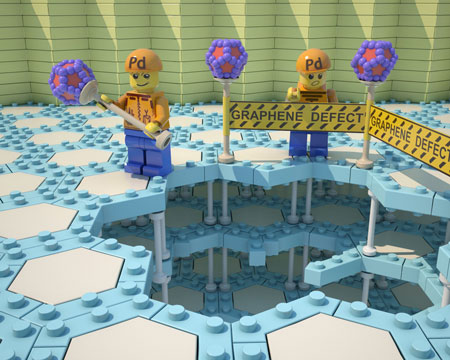
concept of imaging of carbon defect areas by palladium markers
Artistic image on the concept of imaging of carbon defect areas by palladium markers. (Graphene defects marked by Pd). (Image : Ananikov Laboratory, Zelinsky Institute of Organic Chemistry)
When the palladium complex Pd2(dba)3 is dissolved in chloroform, it forms a dark red solution under normal circumstances. But when graphene or another carbon material is added to the solution, the palladium is completely consumed. As a result, the solution turns from dark red to colourless.
Using advanced imaging techniques, the researchers found that the palladium clusters selectively attach to graphene’s surface according to specific patterns, depending on how reactive the carbon centres are. Individual palladium particles settle onto point defects, local accumulations of particles are present on larger defects, and short chains outline linear defects.
These defects are normally invisible under an electron microscope. The palladium particles act like a contrast agent, allowing the spatial imaging of the chemical reactivity, and thus the defects, of graphene layers.
“Metal mapping of carbon materials provides unique insights and reveals hidden information about fascinating properties at the molecular level,” says project leader Professor Valentine Ananikov.
The team’s findings indicate that using palladium markers, more than 2,000 surface defects, or reactivity centres, on graphene can be individually located, per square micrometre of surface area. The researchers say that the unexpected capacity of graphene to accommodate so many reactivity centres challenges scientists to re-examine their understanding of the electronic and structural properties of carbon materials.
Now that the researchers have learned how to recognise and characterise the defects, their next step is to develop a technique to control them. Some defects possess a dynamic nature and have the ability to “migrate” over graphene’s surface. If the researchers can control this migration, they will have a unique opportunity to form materials with customised properties. This is an outstanding direction for future studies, they say
Source: Zelinsky Institute of Organic Chemistry
Subscribe to a free copy of one of our daily
Nanowerk Newsletter Email Digests
with a compilation of all of the day’s news.

Read more: New technique allows to visualize defects on the surface of graphene

Corpurile rapide, elastice ar putea da naștere unui nou val de electronice portabile

Înlătură termen: dimensiuni ale lungimii de undă cuprinse între un milimetru și un metru dimensiuni ale lungimii de undă cuprinse între un milimetru și un metruÎnlătură termen: frecvențele radio cu microunde sunt unde electromagnetice care utilizează frecvențe în intervalul de la 3 gigahertzi la 300 gigahertzi. Aceasta se încadrează direct în domeniul 5G frecvențele radio cu microunde sunt unde electromagnetice care utilizează frecvențe în intervalul de la 3 gigahertzi la 300 gigahertzi. Aceasta se încadrează direct în domeniul 5GÎnlătură termen: noile circuite integrate ar putea fi utilizate în electronice portabile care aderă la piele noile circuite integrate ar putea fi utilizate în electronice portabile care aderă la piele

Posted: May 30, 2016

Fast, stretchy circuits could yield new wave of wearable electronics

(Nanowerk News) The consumer marketplace is flooded with a lively assortment of smart wearable electronics that do everything from monitor vital signs, fitness or sun exposure to play music, charge other electronics or even purify the air around you – all wirelessly.

Now, a team of University of Wisconsin-Madison engineers has created the world’s fastest stretchable, wearable integrated circuits, an advance that could drive the Internet of Things and a much more connected, high-speed wireless world.
Led by Zhenqiang „Jack” Ma, the Lynn H. Matthias Professor in Engineering and Vilas Distinguished Achievement Professor in electrical and computer engineering at UW-Madison, the researchers published details of these powerful, highly efficient integrated circuits in the journal Advanced Functional Materials („Stretchable Twisted, Pair Transmission Lines for Microwave Frequency Wearable Electronics”).
Stretchy Circuit
Fabricated in interlocking segments like a 3-D puzzle, the new integrated circuits could be used in wearable electronics that adhere to the skin like temporary tattoos. Because the circuits increase wireless speed, these systems could allow health care staff to monitor patients remotely, without the use of cables and cords. (Image: Yei Hwan Jung and Juhwan Lee/University of Wisconsin-Madison)
The advance is a platform for manufacturers seeking to expand the capabilities and applications of wearable electronics – including those with biomedical applications – particularly as they strive to develop devices that take advantage of a new generation of wireless broadband technologies referred to as 5G.
With wavelength sizes between a millimeter and a meter, microwave radio frequencies are electromagnetic waves that use frequencies in the .3 gigahertz to 300 gigahertz range. That falls directly in the 5G range.
In mobile communications, the wide microwave radio frequencies of 5G networks will accommodate a growing number of cellphone users and notable increases in data speeds and coverage areas.
In an intensive care unit, epidermal electronic systems (electronics that adhere to the skin like temporary tattoos) could allow health care staff to monitor patients remotely and wirelessly, increasing patient comfort by decreasing the customary tangle of cables and wires.
What makes the new, stretchable integrated circuits so powerful is their unique structure, inspired by twisted-pair telephone cables. They contain, essentially, two ultra-tiny intertwining power transmission lines in repeating S-curves.
This serpentine shape – formed in two layers with segmented metal blocks, like a 3-D puzzle – gives the transmission lines the ability to stretch without affecting their performance. It also helps shield the lines from outside interference and, at the same time, confine the electromagnetic waves flowing through them, almost completely eliminating current loss. Currently, the researchers’ stretchable integrated circuits can operate at radio frequency levels up to 40 gigahertz.
And, unlike other stretchable transmission lines, whose widths can approach 640 micrometers (or .64 millimeters), the researchers’ new stretchable integrated circuits are just 25 micrometers (or .025 millimeters) thick. That’s tiny enough to be highly effective in epidermal electronic systems, among many other applications.
Ma’s group has been developing what are known as transistor active devices for the past decade. This latest advance marries the researchers’ expertise in both high-frequency and flexible electronics.
„We’ve found a way to integrate high-frequency active transistors into a useful circuit that can be wireless,” says Ma, whose work was supported by the Air Force Office of Scientific Research. „This is a platform. This opens the door to lots of new capabilities.”
Source: By Renee Meiller, University of Wisconsin-Madison

Read more: Fast, stretchy circuits could yield new wave of wearable electronics

Utilizarea nanomaterialelor și produselor nanometrice pentru tratarea apelor uzate(Use of nanomaterials and nanoscale products for wastewater treatment)

Procesele existente de apă și deșeuri Tratarea apei poate fi optimizat prin aplicații adecvate nanotehnologice, care poate duce la o îndepărtare mai bine direcționată a contaminanți, o mai bună eficiență energetică și costuri mai mici.
Experții prezic au, prin urmare, sectorul de apă ar putea beneficia cel mai mult de aplicații nanotehnologice inovatoare în termen mediu și lung

Posted: May 30, 2016

Use of nanomaterials and nanoscale products for wastewater treatment

(Nanowerk News) The German Environment Agency (UBA) has published the factsheet „Use of nanomaterials and nanoscale products for wastewater treatment” (pdf).

The treatment of contaminated water prior to its introduction into surface water is one of the most pressing global challenges of the future.
Existing processes for treating water and wastewater can be optimized by suitable nanotechnology applications, which can result in a more targeted removal of contaminants, better energy efficiency and lower costs.
Experts predict that the water sector could also benefit most from innovative nanotechnology applications in the medium to long term.
In addition to the benefits of using nanomaterials for water treatment, it should also be borne in mind that nanomaterials can have unintended impacts on the environment and human health.
When using nanomaterials it is important to note that the novel properties of nanomaterials may possibly also have harmful effects on the environment and humans.
Additional relevant publications:
Use of nanoscale iron for the remediation of groundwater damages
Use of nanomaterials in coatings
Use of nanomaterials in textiles
Nanotechnology-based lighting systems: organic light-emitting diode (OLED)
Source: German Environment Agency

Read more: Use of nanomaterials and nanoscale products for wastewater treatment

Grupul NanoSafety publică a doua ediție a ghidului pentru a sprijini munca în condiții de siguranță cu nanomaterialele

Este aplicabil unei game largi de nanomateriale, Inclusiv particule, fibre, pulberi, tuburi și fire precum și agregate și aglomeratului, și recunoaște incertitudinea anterioare și actuale în dezvoltarea de gestionare eficientă a riscurilor În cazul în care se ocupă cu nanomaterialele și pledează pentru o strategie de precauție pentru a minimiza potențialul expunere.

Posted: May 30, 2016

NanoSafety Group publishes 2nd Edition of guidance to support safe working with nanomaterials

(Nanowerk News) The UK Nanosafety Group (UKNSG) has updated and published a 2nd edition of guidance (pdf) to support safe and responsible working practices with nanomaterials in research and development laboratories.

Working Safely with Nanomaterials in Research and Development
The document aims to provide guidance on factors relating to establishing a safe workplace and good safety practice when working with particulate nanomaterials.It is applicable to a wide range of nanomaterials, including particles, fibres, powders, tubes and wires as well as aggregates and agglomerates, and recognises previous and current uncertainty in developing effective risk management when dealing with nanomaterials and advocates a precautionary strategy to minimise potential exposure.

The 2nd edition of the guidance provides updates to account for changes in legislation, recent studies in the literature, and best practice since 2012.

In particular, specific sections have been revised to account for the full implementation of Global Harmonised System (GHS) which came into force on 1 June 2015 through the CLP regulations. The document explains the approaches that are presently being used to select effective control measures for the management of nanomaterials, more specifically control banding tools presently in use. Significant changes can be found in the following sections: ‘Hazard Banding’, ‘Exposure Control’, ‘Toxicology’, and ‘Monitoring’.

Of relevance to employers, managers, health and safety advisors, and users of particulate nanomaterials in research and development, the guidance should be read in conjunction with the Approved Code of Practice on COSHH, together with the other literature referred to in the document.
The document has been produced taking account of the safety information currently available and is presented in the format of guidance and recommendations to support implementation of suitable protocols and control measures by employers and employees. It is intended that the document will be reviewed and updated on a periodic basis to keep abreast of the evolving nature of the content.
Source: UK Nanosafety Group

Read more: NanoSafety Group publishes 2nd Edition of guidance to support safe working with nanomaterials

Sisteme de viteze tactice avansate cu acționare directă Spitfire Modulară Armă tactilă Plate Carrier echipată cu acțiune directă Hurricane Rug / Rifle Mag (Magazine) Carrier / Pouch Panel în Hyde Definiție Pencott-GreenZone Foliage Green Combat Camo (camuflaj) Pattern(Direct Action Advanced Tactical Gear Systems Spitfire Modular Tactical Armor Plate Carrier Outfitted with Direct Action Hurricane Chest Rig/Rifle Mag(Magazine) Carrier/Pouch Panel in Hyde Definition PenCott-GreenZone Foliage Green Combat Camo(Camouflage) Pattern)

Abordarea sistemelor Direct Action pentru combaterea / designul tactic al angrenajelor este evidentă în mod special în transportatorul de plăci modulare DA Spitfire, care poate fi configurat în numeroase moduri, în funcție de preferința operatorului

Direct

Acțiune directă Advanced Tactical Gear Systems Combate Imbracaminte / Battle Uniforma (BDU) Prototypes: Prototype Combat tricou și pantaloni(Direct Action Advanced Tactical Gear Systems Combat Clothing/Battle Dress Uniform(BDU) Prototypes:Prototype Combat Shirt and Pants )

Combat tricou și pantaloni;Totul este conceput să facă parte dintr-un SISTEM, pentru a optimiza performanța atacatorului / operatorului

Direct Action

Cercetătorii au controlat eliberarea de proteine ​​din nanoparticule fără încapsulare

De zeci de ani, inginerii biomedicale au fost încapsularea cu migală de proteine în nanoparticule pentru a controla eliberarea lor. Acum, o echipa de cercetare condusa de profesorul universitar Molly Shoichet a demonstrat a proteinelor poate fi lansat peste cateva saptamani, chiar luni, fără a mai fi încapsulate. În acest caz, echipa de analizat în mod special proteine terapeutice relevante pentru regenerarea tesutului dupa accident vascular cerebral și leziuni ale măduvei spinării.

Researchers control protein release from nanoparticles without encapsulation

(Nanowerk News) A University of Toronto Engineering team has designed a simpler way to keep therapeutic proteins where they are needed for long periods of time. The discovery is a potential game-changer for the treatment of chronic illnesses or injuries that often require multiple injections or daily pills.

For decades, biomedical engineers have been painstakingly encapsulating proteins in nanoparticles to control their release. Now, a research team led by University Professor Molly Shoichet has shown that proteins can be released over several weeks, even months, without ever being encapsulated. In this case the team looked specifically at therapeutic proteins relevant to tissue regeneration after stroke and spinal cord injury.
„It was such a surprising and unexpected discovery,” said co-lead author Dr. Irja Elliott Donaghue, who first found that the therapeutic protein NT3, a factor that promotes the growth of nerve cells, was slowly released when just mixed into a Jello-like substance that also contained nanoparticles. „Our first thought was, ‘What could be happening to cause this?'”
Left to right: Irja Elliott Donaghue, Malgosia Pakulska and Jaclyn Obermeyer
Left to right: Irja Elliott Donaghue, Malgosia Pakulska and Jaclyn Obermeyer, under the direction of Professor Molly Shoichet in U of T Engineering, discovered that proteins can be released slowly without first encapsulating them in nanoparticles.
Proteins hold enormous promise to treat chronic conditions and irreversible injuries – for example, human growth hormone is encapsulated in these tiny polymeric particles, and used to treat children with stunted growth. In order to avoid repeated injections or daily pills, researchers use complicated strategies both to deliver proteins to their site of action, and to ensure they’re released over a long enough period of time to have a beneficial effect.
This has long been a major challenge for protein-based therapies, especially because proteins are large and often fragile molecules. Until now, investigators have been treating proteins the same way as small drug molecules and encapsulating them in polymeric nanoparticles, often made of a material called poly(lactic-co-glycolic acid) or PLGA.
As the nanoparticles break down, the drug molecules escape. The same process is true for proteins; however, the encapsulating process itself often damages or denatures some of the encapsulated proteins, rendering them useless for treatment. Skipping encapsulation altogether means fewer denatured proteins, making for more consistent protein therapeutics that are easier to make and store.
„This is really exciting from a translational perspective,” said Jaclyn Obermeyer. „Having a simpler, more reliable fabrication process leaves less room for complications with scale-up for clinical use.”
The three lead authors, Elliott Donoghue, Obermeyer and Dr. Malgosia Pakulska have shown that to get the desired controlled release, proteins only need to be alongside the PLGA nanoparticles, not inside them. Their work was published today in the journal Science Advances („Encapsulation-free controlled release: Electrostatic adsorption eliminates the need for protein encapsulation in PLGA nanoparticles”).
„We think that this could speed up the path for protein-based drugs to get to the clinic,” said Elliott Donaghue.
The mechanism for this encapsulation-free controlled release is surprisingly elegant. Shoichet’s group mixes the proteins and nanoparticles in a Jello-like substance called a hydrogel, which keeps them localized when injected at the site of injury. The positively charged proteins and negatively charged nanoparticles naturally stick together. As the nanoparticles break down they make the solution more acidic, weakening the attraction and letting the proteins break free.
„We are particularly excited to show long-term, controlled protein release by simply controlling the electrostatic interactions between proteins and polymeric nanobeads,” said Shoichet. „By manipulating the pH of the solution, the size and number of nanoparticles, we can control release of bioactive proteins. This has already changed and simplified the protein release strategies that we are pursuing in pre-clinical models of disease in the brain and spinal cord.”
„We’ve learned how to control this simple phenomena,” Pakulska said. „Our next question is whether we can do the opposite–design a similar release system for positively charged nanoparticles and negatively charged proteins.”
Source: University of Toronto

Read more: Researchers control protein release from nanoparticles without encapsulation

Fizica spinului cu ioni prinsi

Ideea propusă de echipa de cercetători arată modul de abordare a problemelor numărului partiționarea prin aplicarea unei strategii cunoscut sub numele de „recoacere cuantică“. Acest lucru se face prin aplicarea unui câmp magnetic puternic INITIALLY Toate care este apoi oprit lent în timpul simulării. În acest fel, starea cuantică este deformată până când se potrivește cu soluția dorită. Acest lucru poate fi mai rapid decât alte metode pentru a rezolva pe problemă.

Posted: May 27, 2016

Spin glass physics with trapped ions

(Nanowerk News) One of the most striking discoveries of quantum information theory is the existence of problems that can be solved in a more efficient way with quantum resources than with any known classical algorithm.

Number-partitioning, which refers to the simple task of dividing a set of numbers into two groups of equal sums is, in fact, a very difficult problem to solve with classical computers.
Often, such problems can be related to a physical model, which then allows for solving the problem by finding the minimum energy state of the model. Here, an important role is played by spin glass models, that is, models describing a collection of tiny magnets interacting with each other in a random fashion.
Solving Number Partitioning with Spin Glass Models
Solving Number partitioning with spin glass models. (Image: ICFO)
In the recent work published in Nature Communications („Quantum annealing for the number-partitioning problem using a tunable spin glass of ions”), Dr. Tobias Grass, David Raventós, Dr. Christian Gogolin, led by ICREA Prof. at ICFO Dr. Maciej Lewenstein, in collaboration with Dr. Bruno Julià-Díaz from the University of Barcelona (UB), lay the theoretical foundations for a quantum simulation of spin glass physics with trapped ions.
The idea proposed by the team of researchers shows how to tackle the problem of number partitioning by applying a strategy known as „quantum annealing”. This is done by initially applying a strong magnetic field which is then switched off slowly during the simulation. In this way, the quantum state is deformed until it matches with the desired solution. This can be faster than other methods to solve the problem.
The implementation of this approach is possible with state-of-the-art techniques for trapping, cooling, and manipulating ions. As Dr. Grass clearly states, „In the past, we have seen quantum simulations which solve a problem from quantum physics. In our approach, the same techniques are used to solve a problem from computer science. The results of our study opens a new path and brings us a step closer to the development of a quantum computer.”
Source: ICFO-The Institute of Photonic Sciences

Read more: Spin glass physics with trapped ions