
Chimiștii combină biologia, nanotehnologia pentru a crea o sursă alternativă de energie

Au demonstrat recent transferul energetic de înaltă eficiență între tijele cuantice semiconductoare și enzimele luciferazăș transfer de energie prin rezonanță bioluminescență

Posted: Feb 29, 2016

Chemists combine biology, nanotechnology to create alternate energy source

(Nanowerk News) Chemists in Syracuse University’s College of Arts and Sciences have made a transformational advance in an alternate lighting source–one that doesn’t require a battery or a plug.

Associate Professor Mathew Maye and a team of researchers from Syracuse, along with collaborators from Connecticut College, have recently demonstrated high-efficient energy transfer between semiconductor quantum rods and luciferase enzymes. Quantum rods and luciferase enzymes are nanomaterials and biomaterials, respectively. When combined correctly, these materials produce bioluminescence–except, instead of coming from a biomaterial, such as a firefly enzyme, the light eminates from a nanomaterial, and is green, orange, red, or near-infrared in color.
A schematic of a nano-biosystem (top) and an electron microscope image of quantum rods
A schematic of the nano-biosystem (top) and an electron microscope image of quantum rods.
The findings are the subject of a recent article in ACS Nano („Probing Bioluminescence Resonance Energy Transfer in Quantum Rod–Luciferase Nanoconjugates”).
„Think of our system as a design project,” Maye says. „Our goal has been to build a nano-biosystem that’s versatile enough to teach us a lot, while allowing us to overcome significant challenges in the field and have practical applications. The design involves materials from our chemistry and biology labs, as well as various nanoscience and self-assembly tools. It’s a true team effort with multiple collaborations.”
Maye illustrates his point by referencing quantum rods, each of which is four nanometers wide and 50 nanometers long. (A nanometer is 1 billionth of a meter.) „The rods were chemically synthesized with amazing precision,” he says. „To get the best information, we realized that we needed at least two different types of rods, each with three synthetically tuned variations, and up to 10 different assembly conditions.”
Having a wide range of variables has enabled Maye and his team to learn more about the science of nano-biology energy transfer.
Prior to becoming a postdoc at the University of Notre Dame, Rabeka Alam G’13 led the project at Syracuse as a Ph.D. student. She says this work illuminates a special kind of interaction known as bioluminescence resonance energy transfer (BRET). „In nanoscience, a quantum dot or rod is typically an energy donor,” she says. „In our case, the energy came from bioluminescent luciferase.”
With BRET, the enzyme is attached to the surface of the rod. Luciferin is added, and acts as a kind of fuel. When the enzyme and fuel interact, they release an energy that is transferred to the rod, causing it to glow.
„The trick to increasing the efficiency [of BRET] is finding the right donor-acceptor combination, which requires different rods and enzymes,” says Liliana Karam, a Syracuse Ph.D. student who currently heads up the project. „Thanks to our colleagues at Connecticut College, we have genetically manipulated enzymes of multiple colors that are attached to the rods, which, in turn, are prepared in our lab at Syracuse.”
Maye says the quantum rods are composed of semi-conductive elements–specifically, an outer shell of cadmium sulfide and an inner core of cadmium selenide. By manipulating the size and shape of the core, the length of the rod, and the way the enzymes are attached and packed onto the surface of the rod, researchers are able to alter the color and intensity of the light that is emitted, thus demonstrating the overall efficiency of the process.
Postodc Tennyson Doane, a senior member of the Maye Research Group, says one of the breakthroughs of the project involves a special type of rod known as a „rod-in-rod.” The group has been hypothesizing why this particular rod results in high-efficiency gains.
„When you have a rod-shaped core, the resulting fluorescence is polarized, meaning circular light comes in, and linearly polarized light comes out,” says Doane, adding that the shape of the material makes BRET more efficient. „We believe that, when aligned correctly with the luciferase-excited state, the rod experiences efficiency gains that otherwise are not witnessed in a self-assembled nanosystem. Controlling the enzyme location and bioluminescence polarization may, one day, lead to novel ‘light switches,” in which only certain enzymes around the quantum rod are able to interact via BRET.”
Maye calls this „using biology for non-biological applications.”
„Our nanorods are made of the same materials used in computer chips, solar panels, and LED [light-emitting diode] lights. At the moment, our system works best in the red to near-infrared range, which has longer wavelengths than visible light, and is invisible to the eye,” he says, alluding to night-vision goggles, medical imaging, and rapid microbial detection. „Our work is patent-pending at Syracuse. Perhaps, we’ll someday have firefly-covered nanorods that can be inserted into LED lights and don’t require a plug.”
Source: Syracuse University

Read more: Chemists combine biology, nanotechnology to create alternate energy source
Interferența la o fantă dublă formată din doi atomi

Emițătoare cu un singur foton utilizând atomi unici stocați în rezonatoare optice ș schimbul de informații cuantice într-o rețea cuantică de lungă distanță.

Posted: Feb 29, 2016

Interference at a double slit made of two atoms

(Nanowerk News) The investigation and exploitation of light-matter-interaction in optical resonators is one of the central research topics in the Quantum Dynamics Division of Professor Gerhard Rempe, Director at the Max Planck Institute of Quantum Optics in Garching. A couple of years ago, the team succeeded in creating single-photon emitters using single atoms stored in optical resonators. The stationary atoms can, for example, serve as nodes for the exchange of quantum information in a long-distance quantum network.

Now, the scientists went one step further. They trapped a pair of atoms with well-defined relative positions in such a resonator and scattered light from this “double slit”. They observed interference phenomena that contradict well-established intuition. These results were enabled by the development of a technique that allows for position control of the atoms with an accuracy well below the wavelength of the scattered light. One motivation for this experiment is to better understand fundamental aspects of cavity quantum electrodynamics.
Furthermore, the technique paves the way for studying new concepts of entanglement generation between quantum bits and thus opens up new perspectives for quantum information processing (Nature Photonics, „Interference and dynamics of light from a distance-controlled atom pair in an optical cavity”).
Resonant laser light is being scattered from two single atoms
Resonant laser light (red arrow) is being scattered from two single atoms. The effects that arise due to interference (here shown in an artist’s view) are determined by the relative spatial phase of the atoms and the interaction with the light mode of the optical resonator (mirrors in grey). Lower left: fluorescence image of two rubidium atoms. (Graphic: Andreas Neuzner, MPQ)
Key element of the experimental set-up is an optical resonator consisting of two highly-reflecting mirrors spaced by 0.5 mm. Inside the cavity, a so-called optical lattice is generated by crossing two retro-reflected laser beams, one oriented orthogonal to and the other along the resonator axis. The resulting light pattern of bright and dark spots resembles a checker board with a period of about half a micrometre. These spots define lattice sites at which the atoms can be trapped and where they are localized to about 25 nanometres.
At first, a couple of rubidium atoms, precooled to very low temperatures, are loaded into the optical lattice. By detecting their fluorescence light via a high-resolution microscope objective, the atoms can be identified as individual light spots. Excess atoms are subsequently removed by individually heating them up with a resonant laser beam, until only a pair of atoms with the desired spacing remains. “This is the “double-slit” from which the resonant laser light, propagating transversally through the resonator, is scattered”, explains Andreas Neuzner, who performed this experiment as part of his doctoral studies.
„Interference can only be observed if the phase relation between the two light sources is fixed“, explains Dr. Stephan Ritter, another scientist on the experiment. “In order to investigate the interference as a function of the phase, we have to know the position of the atoms with a precision well below the wavelength of 780 nanometres.“ Although the resolution of the imaging system limits the size of the atom images to 1.3 micrometres, the scientists can localize the emitting atoms with an accuracy of 70 nanometres and can thereby assign their position to a particular lattice site. Therefore, the distance between two atoms, typically about 10 micrometres, is precisely known.
The resonator favours emission along its axis and enhances the interaction between the atoms and the scattered light, which is reflected multiple times between the mirrors. The light power leaking through one of the mirrors – i.e. the photon rate – is recorded as a function of the relative phase of the two atoms.
The observed interference pattern displays several intriguing features that are not expected in the simpler picture of two classical dipoles in free space. First, in the case of in-phase (constructive) interference, the intensity is only a factor of 1.3 larger than the rate observed for a single atom, whereas a fourfold larger signal is expected for the simpler picture. This phenomenon goes back to the various light fields inside the resonator that have to be taken into account. In contrast to the classical double-slit experiment, not only the phase relation between the scattered light waves matters. It is rather the superposition of the scattered light with the light field of the resonator that in the end leads to an intensity reduction in the field maxima.
The second feature occurs for out-of-phase (destructive) interference. Here, the photon rate drops below the value measured for a single atom, but does not go to zero as one would expect intuitively. Strikingly, extremely strong intensity fluctuations are observed, so-called photon bunching. “This phenomenon arises, because in the case of destructive interference, the atoms can emit photons only pairwise and at the same time into the resonator”, explains Andreas Neuzner.
„In this experiment we have combined three key techniques for the first time: Using an optical lattice, we position the atoms with high accuracy and then localize them with a high-resolution microscope. The interaction with the resonator enables directed detection of the scattered light“, says Stephan Ritter. “The newly developed techniques are essential for future experiments aiming to explore collective radiation effects predicted for multi-atom systems“, resumes Prof. Gerhard Rempe. “On the other hand, they offer the possibility to implement novel protocols for quantum information processing with several quantum bits.”
Source: Max Planck Institute of Quantum Optics

Read more: Interference at a double slit made of two atoms

Dispozitive electronice extensibile care au o lungime cvadruple(Stretchable electronics that quadruple in length)

Și pentru că urmează forma și mișcarea corpului uman, ar putea fi folosită pentru senzori concepuți pentru a monitoriza anumite funcții biologice

sti (6)

Controlul artificial al exciplexurilor deschide posibilități pentru noile electronice

Posted: Feb 26, 2016

Artificial control of exciplexes opens possibilities for new electronics

(Nanowerk News) Demonstrating a strategy that could form the basis for a new class of electronic devices with uniquely tunable properties, researchers at Kyushu University were able to widely vary the emission color and efficiency of organic light-emitting diodes based on exciplexes simply by changing the distance between key molecules in the devices by a few nanometers. This new way to control electrical properties by slightly changing the device thickness instead of the materials could lead to new kinds of organic electronic devices with switching behavior or light emission that reacts to external factors.

Organic electronic devices such as OLEDs and organic solar cells use thin films of organic molecules for the electrically active materials, making flexible and low-cost devices possible.
A key factor determining the properties of organic devices is the behavior of packets of electrical energy called excitons. An exciton consists of a negative electron attracted to a positive hole, which can be thought of as a missing electron.
In OLEDs, the energy in these excitons is released as light when the electron loses energy and fills the vacancy of the hole. Varying the exciton energy, for example, will change the emission color.
However, excitons are commonly localized on a single organic molecule and tightly bound with binding energies of about 0.5 eV. Thus, entirely new molecules must usually be designed and synthesized to obtain different properties from these Frenkel-type excitons, such as red, green, or blue emission for displays.
Researchers at Kyushu University’s Center for Organic Photonics and Electronics Research (OPERA) instead focused on a different type of exciton called an exciplex, which is formed by a hole and electron located on two different molecules instead of the same molecule.
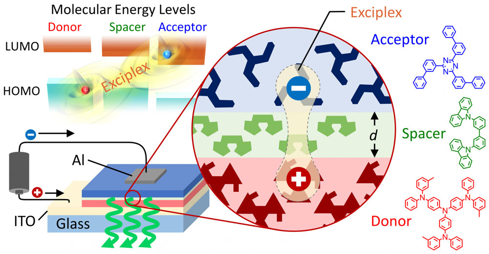
By manipulating the molecular distance between the electron-donating molecule (donor) and the electron-accepting molecule (acceptor) that carry the exciplex’s hole and electron, respectively, the researchers could modify the properties of these weakly bound excitons.
basic structure of an exciplex-based OLED with emission color and efficiency that can be controlled simply by changing the spacer thickness
This schematic shows the basic structure of an exciplex-based OLED with emission color and efficiency that can be controlled simply by changing the spacer thickness. An exciplex forms when a hole in the highest occupied molecular orbital (HOMO) of a donor molecule is attracted to an electron in the lowest unoccupied molecular orbital (LUMO) of an acceptor molecule. Light is emitted when the electron releases energy as light and transfers across the spacer layer to the donor molecule, thus replacing the missing electron represented by the hole. The thickness of the spacer can be used to modify the attraction between the hole and electron and tune the exciplex energy. Without changing any of the molecules, emission color could be varied from orange to yellowish green and the efficiency enhanced eight fold by increasing the spacer thickness to 5 nm. (click on image to enlarge)
„What we did is similar to placing sheets of paper between a magnet and a refrigerator,” said Associate Professor Hajime Nakanotani, lead author of the paper reporting these results published online February 26, 2016, in the journal Science Advances („Long-range coupling of electron-hole pairs in spatially separated organic donor-acceptor layers”).
„By increasing the thickness of an extremely thin layer of organic molecules inserted as a spacer between the donor and acceptor, we could reduce the attraction between the hole and electron in the exciplex and thereby greatly influence the exciplex’s energy, lifetime, and emission color and efficiency.”
Indeed, the changes can be large: by inserting a spacer layer with a thickness of only 5 nm between a donor layer and an acceptor layer in an OLED, the emission color shifted from orange to yellowish green and the light emission efficiency increased 700%.
For this to work, the organic molecule used for the spacer layer must have an excitation energy higher than those of the donor and acceptor, but such materials are already widely available.
While the molecular distance is currently determined by the thickness of the vacuum-deposited spacer layer, the researchers are now looking into other ways to control the distance.
„This gives us a powerful way to greatly vary device properties without redesigning or changing any of the materials,” said Professor Chihaya Adachi, director of OPERA. „In the future, we envision new types of exciton-based devices that respond to external forces like pressure to control the distance and electrical behavior.”
In addition, the researchers found that the exciplexes were still formed when the spacer was 10 nm thick, which is long on a molecular scale.
„This is some of the first evidence that electrons and holes could still interact like this across such a long distance,” commented Professor Adachi, „so this structure may also be a useful tool for studying and understanding the physics of excitons to design better OLEDs and organic solar cells in the future.”
„From both scientific and applications standpoints, we are excited to see where this new path for exciton engineering takes us and hope to establish a new category of exciton-based electronics.”
Source: Kyushu University

Read more: Artificial control of exciplexes opens possibilities for new electronics

Determinarea structurilor produselor farmaceutice nanocristaline prin difracție electronică

Posted: Feb 26, 2016

Determining the structures of nanocrystalline pharmaceuticals by electron diffraction

(Nanowerk News) Reliable information about the structure of pharmaceutical compounds is important for patient safety, for the development of related drugs and for patenting purposes. However, working out the structures of pharmaceuticals can be tough.

90° cut-off schematic of the camera as designed for the CM200
90° cut-off schematic of the camera as designed for the CM200 (Technische Universiteit Delft, The Netherlands).
The individual molecules can pack together in the solid in different ways to form different polymorphs, and pertinent properties such as stability, bioavailability or how fast they dissolve in the stomach can vary from one polymorph to another. Single crystals (as used in standard X-ray diffraction experiments) therefore might not be representative of the bulk sample, or indeed might not even be available.

Moreover, the compounds themselves can be damaged by the high energy of the X-radiation used. As electrons are less damaging than X-rays by several orders of magnitude, using electron diffraction should be an attractive alternative, particularly when only nanometre-sized crystals are available. Cooling the sample to liquid-nitrogen temperatures (‘cryo-cooling’) can also help to minimize radiation damage, but the compound might change structure on cooling, so the structure that is obtained is not actually that of the material as taken by the patient at room temperature.

A group of scientists from a number of European countries have tackled all aspects of these problems by using low-dose electron diffraction, rotating the sample so that individual nanocrystals are not in the electron beam long enough to be damaged and collecting the diffraction data using a new type of detector developed by CERN („Ab initio structure determination of nanocrystals of organic pharmaceutical compounds by electron diffraction at room temperature using a Timepix quantum area direct electron detector”).

This new detector combines a high dynamic range with a very high signal-to-noise ratio and sensitivity to single electrons. Radiation damage was reduced so much that cooling the sample was not found to be necessary, allowing the team to study the anticonvulsant drug carbamazepine and nicotinic acid (vitamin B3) at room temperature. The data they collected were high enough quality that they could solve the structures of the two compounds using direct methods and software developed for X-ray crystallography.
Based on their experience with these case studies, the authors are planning to improve the design of their experimental setup further, and will also be developing programs specifically designed for handling electron-diffraction data.
Source: International Union of Crystallography

Read more: Determining the structures of nanocrystalline pharmaceuticals by electron diffraction

Cum cresc grupurile metalice

Procesele de transformare a clusterelor metalice au loc rapid. În mod normal, nu este posibil să se respecte aceste procese și produsele intermediare. „Chiar și primii pași sunt în mare parte necunoscuți și pot fi identificați numai prin combinarea sintezei cu măsurarea și modelarea chimică computerizată”, spun experții. Dacă s-ar cunoaște toate etapele de dezvoltare, grupurile metalice cu proprietăți optoelectronice și magnetice ajustate fin pot fi personalizate pentru aplicații tehnice.
În studiul publicat acum, echipa raportează formarea unui cluster metalic multinucleat, prin sintetizarea mai întâi a unei serii de clustere cu dimensiuni variabile ale metaloizilor germaniu și arsenic. Clusterele mai mari au un atom de tantal de metal tranzițional în centrul moleculelor de colivie. Măsurătorile și simulările pe calculator sugerează că metalul de tranziție joacă foarte devreme un rol în formarea clusterelor.

Posted: Feb 26, 2016

How metal clusters grow

(Nanowerk News) First the nucleus, then the shell: Researchers from Marburg and Karlsruhe have studied stepwise formation of metal clusters, smallest fractions of metals in molecular form. The shell gradually forms around the inner atom rather than by later inclusion of the central atom. Knowledge of all development steps may allow for customized optoelectronic and magnetic properties, as is reported by the researchers in the science journal Nature Communications („Understanding of multimetallic cluster growth”).

formation of a metal cluster from the atomic constituents to the compound
Step by step towards the complete shell: formation of a metal cluster from the atomic constituents to the compound. (Image: Dehnen Group, Philipps-Universität Marburg) (click on image to enlarge)
To specifically synthesize chemical compounds, mechanisms responsible for their formation have to be known. „Purely inorganic compounds are a black box in this respect,” Florian Weigend of Karlsruhe Institute of Technology (KIT) and Stefanie Dehnen of Philipps-Universität Marburg, the corresponding authors of this study, explain. „This particularly applies to the formation of multinucleated metal complexes, so-called clusters.”
Processes of transforming metal clusters take place rapidly. Normally, it is not possible to observe these processes and the intermediate products. „Even the very first steps are largely unknown and can only be identified by combining synthesis with measurement and computerized chemical modeling,” the experts say. If all development steps were known, metal clusters with finely adjusted optoelectronic and magnetic properties might be customized for technical applications.
In the study published now, the team reports the formation of a multinucleated metal cluster by first synthesizing a series of variably sized clusters of the metalloids germanium and arsenic. Larger clusters have an atom of the transition metal tantalum in the center of the cage molecules. Measurements and computer simulations suggest that the transition metal very early plays a role in cluster formation.
„It may be considered a type of catalyst that initiates the formation and breaking of bonds,” Weigend and Dehnen point out. Altogether, the findings show that the transition metal does not settle into an earlier formed cluster shell, but that the shell of the cluster gradually forms around the atom in the center. „The results can be generalized for a whole family of metallic cluster compounds,” Weigend and Dehnen say.
Apart from chemical syntheses and measurements in Marburg, extensive computer simulations with the largely KIT-developed quantum chemistry program package TURBOMOLE were made at KIT. In this way, the role of the many isomers in the reaction was determined. Isomers are chemical compounds of identical composition, but with different spatial arrangements of the atoms.
„Thanks to the calculation of reaction paths, we have found that transformations of isomers are possible with a relatively small energy expenditure,” Weigend says.
Source: Karlsruhe Institute of Technology

Read more: How metal clusters grow

Un nou dispozitiv ar putea pune capăt agoniei pentru mii de oameni(New device could end blood test agony for thousands)

a creat un senzor productiv în masă, care poate detecta nivelele de amoniac în respirație – o descoperire care ar putea elimina utilizarea acelor în rândul a mii de pacienți

s40

Robotul auto-direcționat poate identifica obiecte(Self-directed robot can identify objects)

sunt un Graal Sfânt pentru oamenii de știință, cu aplicații potențiale care includ explorarea peisajelor planetelor îndepărtate
Acestea sunt făcute cu un coș de trepte de roți, înălțate de două picioare, care se rostogolesc în jurul podelei și care își proclamă descoperirile ca fiind „un ochi”, o cameră atașată, cade pe ele

s38

Șocant roz: un test inexpresiv pentru atacurile cu arme chimice(Shocking Pink: An Inexpresive Test for Chemical Weapon Attacks)

ar putea inspira oamenii de stiinta pentru a dezvolta un instrument de cost pentru a detecta rapid arme chimice in aer; au fost în măsură să detecteze prezența unui agent nervos legat de gazul sarin la un nivel scăzut

20 sept